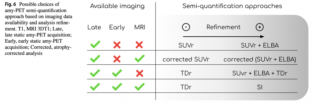

To date, there is no consensus on how to semi-quantitatively assess brain amyloid PET.
Researchers used data from 85 patients who underwent dual time-point PET/MRI acquisitions. The correlations with a gold standard (SI) were computed and the methods compared with the visual assessment.
Each quantifier exhibited excellent agreement with visual assessment and strong correlation with SI (average AUC = 0.99, ρ = 0.91). Among the other methods, TDr came closest to the reference with less implementation complexity.
The ability of techniques integrating blood perfusion to depict age-related variations in amyloid load in amyloid-negative subjects demonstrates the goodness of the estimate.

Peira E, Poggiali D, Pardini M, Barthel H, Sabri O, Morbelli S, Cagnin A, Chincarini A, Cecchin D. A comparison of advanced semi-quantitative amyloid PET analysis methods. Eur J Nucl Med Mol Imaging. 2022 Jun 2. doi: 10.1007/s00259-022-05846-1. Epub ahead of print. PMID: 35652962.


