Melone M, Ciriachi C, Pietrobon D, Conti F
Cereb Cortex. 2019 Jul 22;29(8):3331-3350 (2019)
Melone M, Ciriachi C, Pietrobon D, Conti F
Cereb Cortex. 2019 Jul 22;29(8):3331-3350 (2019)
Iure A, Mazzocchetti P, Bastioli G, Picconi B, Costa C, Marchionni I, Casari G, Tozzi A, Pietrobon D, Calabresi P
Cephalalgia. 2019 Sep;39(10):1333-1338 (2019)
Di Stefano V, Rispoli MG, Pellegrino N, Graziosi A, Rotondo E, Napoli C, Pietrobon D, Brighina F, Parisi P. J
Neurol Neurosurg Psychiatry. 2020 Jul;91(7):764-771 (2020)
Romanos J, Benke D, Pietrobon D, Zeilhofer HU, Santello M.
Sci Adv. 2020 Jun 5;6(23):eaaz1584 (2020)

by prof. Fritjof Helmchen, Brain Research Institute of the University of Zurich, Switzerland
When: Apr 29th, 2021 – 3:00 pm
Where: Zoom meeting
Abstract: Through the combination of in vivo optical imaging and chronic expression of genetically encoded calcium indicators it is now feasible to directly ‘watch’ brain activity patterns related to specific behaviors. I will introduce wide-field calcium imaging and multi-fiber photometry as two complementary methods that enable measurements across mouse neocortex and in large sets of subcortical regions, respectively. Specific patterns of brain-wide signal flow that occur in mice performing whisker-based or auditory sensory discrimination tasks will be discussed, highlighting salient patterns related to short-term memory. In addition, taking advantage of chronic measurements over weeks, I will present salient changes in brain dynamics that relate to task learning.
Short bio: Fritjof Helmchen received his Diploma in Physics from the University of Heidelberg. He completed his PhD thesis in Neuroscience at the May-Planck-Institute for Medical Research in Heidelberg and received his doctorate from the University of Göttingen in 1996. As a postdoc, Dr. Helmchen worked at the Bell Laboratories, Lucent Technologies, NJ, where he pioneered in vivo applications of two-photon microscopy. He then returned to the Max-Planck-Institute for Medical Research, Heidelberg, heading a junior research group from 2000-2005. In 2005 Dr. Helmchen was appointed Professor of Neurophysiology and Co-Director at the Brain Research Institute of the University of Zurich, Switzerland. Dr. Helmchen’s research is centered on the further development and application of imaging techniques for the study of neural network dynamics and neural computations as the basis of animal perception and behaviour.
Avviso di indagine di mercato
Indagine di mercato finalizzata all’affidamento diretto tramite mercato elettronico (MEPA) ai sensi dell’art. 1, comma 2,lett. a) del D.L. 76/2020 (convertito con Legge 11 settembre 2020, n. 120) della fornitura di un Elettroencefalografo (EEG) e di un Eye-Tracker portatili ed integrati in modo che sia possibile la sincronizzazione del segnale EEG con quello dell’Eye-Tracker per l’Universita’ degli Studi di Padova – Centro di Ateneo Padova Neuroscience Center (PNC) – CUP C24I19002710006
Scadenza per la presentazione delle istanze è il 30 aprile 2021 alle ore 12:00

One position for a postdoctoral fellowship has been opened (see details below) in the context of the following project:
Title of the project
In vivo functional characterization of whole brain- and population-level dynamics in genetic mouse models of migraine.
Abstract of the project
Migraine is a remarkably disabling and still poorly understood brain disorder which primarily affects the sensory nervous system. It is characterized by recurrent attacks of unilateral headache and by a global dysfunction in multisensory information processing. Previous investigations at the microcircuit level in genetic models of migraine have revealed significant alterations in the cortical excitatory synaptic transmission due to impaired mechanisms of glutamate release and glutamate clearance. Goal of the proposed project is investigating, in awake animal models, the impact that these microcircuit-level alterations have on the population and brain wide neuronal dynamics. In particular, the investigation will focus on the characterization of the functional connectivity in resting-state condition and of the spatio-temporal features of the activity elicited by a sensory stimulation. At this purpose, the research will adopt mostly optogenetic approaches based on fluorescence-based genetically encoded reporters of the neuronal activity and reporters of glutamate concentrations (iGluSnFr), in combination with large field of view high-speed mesoscale imaging and multiphoton imaging. To characterize the effective connectivity in the FHM models, in a parallel approach, the research will use light-based activity modulators (ChR2/GtAcR) in combination with activity recording.
Qualifications
We are looking for motivated and highly committed candidates holding a Ph.D in biomedical, biological or biophysical sciences or other related disciplines, or alternatively a minimum of three years of experience with research in mouse models. Ideal candidate should have a proven experience in techniques for research with mouse models in vivo and for functional imaging or optogenetics in vivo. High levels of flexibility and proven ability to manage a research project are a plus.

Webinar, Università di Padova
When: Mar 19th, 2021 – 6:00 pm
Where: Zoom Meeting and live on Facebook
Abstract: La “Brain awareness week” è una ricorrenza annuale dedicata a sollecitare la pubblica consapevolezza nei confronti della ricerca sul cervello. All’iniziativa, coordinata dalla Dana Foundation, partecipano società neuroscientifiche, università, istituti ed enti di ricerca di tutto il mondo.
L’Università di Padova in occasione della Brain Awareness Week, la settimana internazionale di divulgazione delle neuroscienze, propone una serie di incontri online: gli interventi sono tenuti da giovani ricercatori e ricercatrici dell’Università di Padova, appartenenti a diversi dipartimenti e accomunati dallo studio del cervello. I relatori accompagnano dunque nell’affascinante mondo dei neuroni, nelle loro interazioni sino ai processi cognitivi alla base del nostro pensiero.

by prof. Richard Wise, Universita’ degli Studi “G.d’Annunzio” di Chieti-Pescara
When: Mar 18th, 2021 – 3:00 pm
Where: Zoom meeting
Abstract: In the last 30 years blood oxygenation level dependent (BOLD) fMRI has taught us a great detail about human brain organisation. However, it only tells us where brain activity is changing without telling us by how much or what the baseline level of activity is. We and others are working on fMRI methods to map the absolute rate of cerebral metabolic oxygen consumption as a marker of the physiological state of brain tissue in health and disease. Development of these methods has resulted in a toolkit for assessing oxygen consumption and cerebrovascular function (including vascular reactivity and arterial compliance) that is yielding interesting results in our pilot clinical studies in epilepsy and multiple sclerosis and in the study of the healthy brain. These approaches have the potential to take fMRI to a new level of quantification of brain function required for experimental medicine and future clinical application.
Short bio: Richard is a physicist who has always worked at the interface of physics and physiology. He specialised in cardiovascular magnetic resonance imaging for his PhD at Cambridge University. In 2000 he changed from imaging the heart to the brain with a move to Oxford University as a post-doctoral research fellow, (Wellcome and MRC research fellowships). From 2006 he developed his research group at Cardiff University Brain Research Imaging Centre with a focus on understanding drug effects on human brain function and looking for ways to quantify brain function using functional MRI approaches, based on sensitivity to blood oxygenation in the human brain. This has led recently to a new magnetic resonance imaging toolkit with the possibility to interrogate, in detail, the function of brain blood vessels and to measure the amount of oxygen fuel that the human brain is using in health and disease. At the end of 2019 he moved to ITAB (Institute of Advanced Biomedical Technologies) and the Department of Neuroscience, Imaging and Clinical Sciences at the University of Chieti-Pescara in Italy, as full professor of applied physics (Professore Ordinario – chiamata diretta).

Accademia dei Lincei e Fondazione “Guido Donegani”, Roma
When: Mar 5th, 2021 – 9:00 am
Where: Zoom meeting (will be published on Mar 5th). Also live on the Accademia’s channel
Abstract: L’Intelligenza Artificiale è una delle tecnologie che sta trasformando la nostra società e molti aspetti della nostra vita quotidiana. Ha già prodotto molti effetti benefici e può essere sorgente di considerevole prosperità economica. Tuttavia, pone problemi riguardanti, in varia misura, l’occupazione, la riservatezza dei dati, la “privacy”, la violazione di valori etici e la fiducia nei risultati. Problematiche simili, con riferimento a problemi occupazionali, di sicurezza e di possibile violazione di valori etici, si pongono anche per quanto riguarda le trasformazioni che avvengono nella società a seguito dello sviluppo, intimamente connesso a quello dell’AI, dei metodi della robotica e dell’automazione della produzione industriale.
In programma l’intervento del prof. Corbetta dal titolo “Il ruolo dell’attività spontanea nell’ architettura funzionale del cervello e nella cognizione”.

by prof. Andrea Crisanti, Molecular Medicine Department, University of Padova
When: March 2nd, 2021 – 5:30 pm
Where: Zoom meeting
To attend the seminar and get the ECM credits you must:
Short bio: Professore di parassitologia molecolare all’Imperial College dal 2000, Andrea Crisanti è attualmente Direttore del Dipartimento di Medicina Molecolare e del Servizio di Microbiologia e Virologia, Azienda Ospedale – Università Padova.
Dopo la laurea in Medicina all’Università di Roma La Sapienza e un dottorato presso l’Istituto di Immunologia di Basilea, Andrea Crisanti ha aperto la strada alla biologia molecolare del vettore della malaria umana Anopheles gambiae contribuendo alla conoscenza genetica e molecolare del parassita della malaria e del suo vettore. Ha ricevuto numerosi riconoscimenti e finanziamenti nazionali e internazionali tra cui Wellcome Trust, BBSRC, la Commissione Europea e NIH.
Autore di numerose pubblicazioni scientifiche di rilievo (oltre 10,000 citazioni, H index 57).
Recentemente parte di una task force scientifica per la gestione dell’emergenza Covid-19 in Veneto.

by prof. Martin Lindquist, School of Public Health, Johns Hopkins University
When: Feb 18th, 2021 – 4:00 pm
Where: Zoom meeting. Recording available on Mediaspace
Abstract: Mediation analysis is an important tool in the behavioral sciences for investigating the role of intermediate variables that lie in the path between a randomized treatment/exposure and an outcome variable. The influence of the intermediate variable on the outcome is often explored using structural equation models (SEMs), with model coefficients interpreted as possible effects. While there has been significant research on the topic in recent years, little work has been done on mediation analysis when the intermediate variable (mediator) is a high-dimensional vector. In this work we introduce a novel method for mediation analysis in this setting called the principal directions of mediation (PDMs). We demonstrate the method using a functional magnetic resonance imaging (fMRI) study of thermal pain where we are interested in determining which brain locations mediate the relationship between the application of a thermal stimulus and self-reported pain.
Short bio: Martin Lindquist is a Professor of Biostatistics at Johns Hopkins University. His research focuses on mathematical and statistical problems relating to functional Magnetic Resonance Imaging (fMRI). Dr. Lindquist is actively involved in developing new analysis methods to enhance our ability to understand brain function using human neuroimaging. He has published over 70 articles, and serves on the editorial boards of several scientific journals both in statistics and neuroimaging. He is a fellow of the American Statistical Association.
In 2018 he was awarded the the Organization for Human Brain Mapping’s ‘Education in Neuroimaging Award’ for teaching statistical issues to the neuroimaging community and the development of online classes that have taught fMRI methods to more than 80,000 students world-wide.

by prof. Olaf Sporns, Department of Psychological and Brain Sciences, Indiana University
When: Jan 28th, 2021 – 3:00 pm
Where: Zoom meeting. Recording available on Mediaspace
Abstract: It is said that complexity lies between order and disorder. In physiology, complexity issues are being considered with increased emphasis. Of crucial importance in the medical setting, pathological activity has been associated with low variability/complexity. In the case of the nervous system, it is well known that excessive synchronization is connected with pathologies such as epilepsy and Parkinson disease. However, brain rhythms and neural synchronization are also crucial for perception and cognition, so it is clear that either too much or not enough synchronization can lead to dysfunctional brain states.
Short bio: After receiving an undergraduate degree in biochemistry, Olaf Sporns earned a PhD in Neuroscience at Rockefeller University and conducted postdoctoral work at The Neurosciences Institute in New York and San Diego. Currently he is the Robert H. Shaffer Chair, a Distinguished Professor, and a Provost Professor in the Department of Psychological and Brain Sciences at Indiana University in Bloomington. Sporns holds adjunct appointments in the School of Informatics, Computing and Engineering, and in the School of Medicine. His main research area is theoretical and computational neuroscience, with a focus on complex brain networks. In addition to over 250 peer-reviewed publications he has written two books, “Networks of the Brain” and “Discovering the Human Connectome”. He is the Founding Editor of “Network Neuroscience”, a journal published by MIT Press. Sporns received a John Simon Guggenheim Memorial Fellowship in 2011, and the Patrick Suppes Prize in Psychology/Neuroscience, awarded by the American Philosophical Society, in 2017. He is a Fellow of the American Association for the Advancement of Science and the Society of Experimental Psychologists and of the Society of Experimental Psychologists.

The “Padova Neuroscience Center-PNC” is glad to inform that one of our PhD Students, Arianna Menardi, has been recently awarded for her presentations during a Conference.

Arianna Menardi won a Poster Award with her presentation “Network-targeted TMS stimulation via individualized target selection: a new route toward enhanced reliability”, during the “Transcranial Brain Stimulation in Cognitive Neuroscience Workshop- II Edition” organized by the Center for Mind/Brain Science- CIMeC (https://event.unitn.it/tbs-cnw/).
The “Padova Neuroscience Center-PNC” is glad to inform that two of our PhD Students, Miriam Celli and Tommaso Volpi, have been recently awarded for their presentations during two Conferences.

Miriam Celli won the “SIPF Youth Award” as Best Scientific Contribution with her presentation, called “Resting-State EEG Signatures of Visual Exploration Styles”, during the XVIII Conference of the Società Italiana di Psicofisiologia e Neuroscienze Cognitive

Tommaso Volpi won the “Gamma Prize 2020” for the Best oral proffered talk, with his presentation called: “The negative relationship between brain metabolism and its network dynamics: stability requires more energy”, during the Pet is Wonderful 2020’s Conference.
“Many animals fall for the same optical illusions we do, providing clues about how evolution shapes visual perception.”
The work of prof. Agrillo has been cited on the National Geographic site

Each year, Clarivate™ identifies the world’s most influential researchers – the select few who have been most frequently cited by their peers over the last decade. In 2020, fewer than 6,200, or about 0.1%, of the world’s researchers, in 21 research fields and across multiple fields, have earned this exclusive distinction.
See the full list here

by prof. Ramón Guevara Erra, DFA – Dept. of Physics and Astronomy – Padova
When: Nov 17th, 2020 – 3:00 pm
Where: Zoom meeting
Abstract: It is said that complexity lies between order and disorder. In physiology, complexity issues are being considered with increased emphasis. Of crucial importance in the medical setting, pathological activity has been associated with low variability/complexity. In the case of the nervous system, it is well known that excessive synchronization is connected with pathologies such as epilepsy and Parkinson disease. However, brain rhythms and neural synchronization are also crucial for perception and cognition, so it is clear that either too much or not enough synchronization can lead to dysfunctional brain states.
Short bio: Ramon Guevara is a physicist at the Department of Physics of the University of Padova. His main interest lies in the interface between biophysics and neuroscience. He has been a research fellow at several institutions, universities and hospitals around the world, including the University Paris Descartes, the Hospital for Sick Children in Toronto, the University of British Columbia in Vancouver and the neuroimage center Neurospin, at Saclay, France. His research focuses on the coordination of neural activity, both in the normal and the pathological brain, and in particular on the search of principles underlying synchronization phenomena in the brain, and their physiological and cognitive implications.

When: December 3rd, 2020 – 3:00 – 5:00 PM CET ( 09:00 – 11:00 AM ET )
Where: Zoom Webinar
Webinar by NEUROMOVE-Rehab Lab, IAS Lab and MAL Lab
Please fill the form to subscribe and receive the link to connect a few days before the Webinar.
Stazi M, Negro S, Megighian A, D’Este G, Solimena M, Jockers R, Lista F, Montecucco C, Rigoni M
J Pineal Res. 2021 Jan;70(1):e12695

When: September 25th, 2020
Where: Aula Ippolito Nievo – Cortile Antico, Palazzo del Bo – Padova
The Padua Neuroscience Center is a unique multidisciplinary environment that integrates many different scientific backgrounds with the common effort to understand the brain mechanisms. In this scenario, sharing scientific ideas and technical expertise represents a crucial and fundamental step to establish collaborative networks and to draft powerful scientific plans.
The PNC Brain Day 2020 is the opportunity for the PIs:

Prof. Carlo Semenza has been recently admitted as a member of the prestigious Academia Europaea.
The object of Academia Europaea is the advancement and propagation of excellence in scholarship in the humanities, law, the economic, social, and political sciences, mathematics, medicine, and all branches of natural and technological sciences anywhere in the world for the public benefit and for the advancement of the education of the public of all ages in the aforesaid subjects in Europe.
Academia Europaea is a European, non-governmental association acting as an Academy. Our members are scientists and scholars who collectively aim to promote learning, education and research. Founded in 1988, with about 3800 members which includes leading experts from the physical sciences and technology, biological sciences and medicine, mathematics, the letters and humanities, social and cognitive sciences, economics and the law.

Decree of acts approval and Ranking
Please note: in order to apply it is compulsory to:
The position
In the context of the European Training Network “European School of Network Neuroscience“ (euSNN), we are offering a full-time 3-year PhD position for a person with a strong background in computational neuroscience and neuroimaging. The successful applicant will be working in Padova at the Padova Neuroscience Center (PNC) and Department of Neuroscience. The position involves the analysis of a unique prospective longitudinal cohort of first-time stroke patients and healthy controls studied at Washington University in St. Louis.
The gross amount per year will be of 39.217,20 € while the net amount per year will be of 34.740 €, family allowance will be granted if necessary.
The candidate will be able to ask the following questions:
1. What are the most common patterns of behavioral deficits post-stroke and the time course of recovery?
2. What are the lesion, structural, functional, and perfusion correlates of acute dysfunction and recovery for different kinds of deficits (motor, visual, attention, memory, emotion, executive)?
3. What is the relationship between resting and task states in patients with focal lesions? How alterations of resting organization reflect task organization?
4. Does anatomical connectivity recover post-stroke?
5. Does post-stroke atrophy play a role in recovery?
6. Do neuroimaging signals allow for a more accurate prediction of outcome?
The candidate will need to apply a variety of statistical models including linear and non-linear methods such as principal component analysis, ridge, lasso and elastic net regression, partial least square regression and correlation, canonical correlation analysis. The candidate will need to learn about basic mechanisms of plasticity and recovery.
The successful applicant will be supervised by Prof. Dr. Maurizio Corbetta, Chair of Neurology and Director of the PNC, and Prof. Alessandra Bertoldo of the Department of Bioengineering and Vice-Director of the PNC. The applicant will work in a multi-disciplinary laboratory with neurologists, psychologists, bioengineers, and physicists.
The early stage researcher (ESR) will be enrolled in the Neuroscience PhD School @PNC in Padova. Furthermore, the ESR will meet and visit partner organizations of the ETN in Europe to deepen and extend knowledge about connectivity measurement and analysis approaches, and to establish a network with other early stage researchers (ESRs) and institutions.
Our ideal candidate
You will hold a master’s degree in computational or cognitive neuroscience, or related fields like biomedical engineering. Your thesis or internships should be related to research with fMRI, computational modelling, or statistics demonstrating relevant expertise and problem-solving skills. Preferably you already have experience with programming in R, Matlab, Python and/or C++ and have experience to run multivariate analysis on neuroimaging datasets. We also expect that you have well developed interpersonal skills to work in a team.
Want to apply?
If this sounds interesting to you, please look at the Competition notice and the Application form and follow the instructions.
The deadline for application is September 30th, 2020 at 13:00 (CET)
Applicants need to meet the European Training Network grant eligibility conditions of ESRs, that are described at this web page:
https://infrastar.eu/recruitment/esr-eligibility-conditions-and-responsabilities/ .
Most relevant is that all nationalities can apply, but only if the applicant did not reside or carried out main activity (such as work or study) in Italy for more than 12 months within the last 3 years.
Zullo L, Bozzo M, Daya A, Di Clemente A, Mancini FP, Megighian A, Nesher N, Röttinger E, Shomrat T, Tiozzo S, Zullo A, Candiani S
Cells. 2020 Aug 19;9(9):1925
(Italian version only) Articolo pubblicato nel sito neurologiaitaliana.it del 17 marzo 2020.
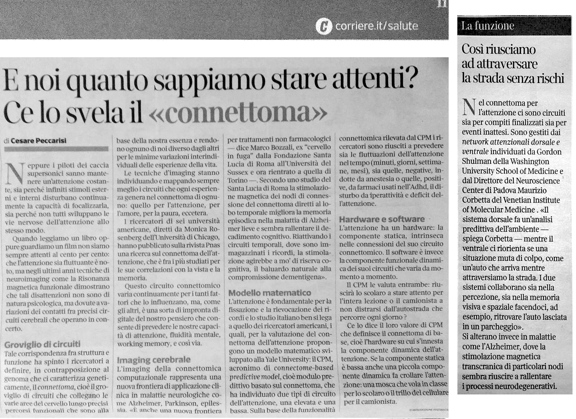
(Italian version only) Articolo pubblicato nell’inserto “Salute” del Corriere della Sera del 16 luglio 2020.
One research grant position has been opened (see details below) in the context of the following project:
Title of the project
Eye movements and brain dynamics in Alzheimer’s disease: the impact of smart natural environments
Interviews date:
August 6th, 2020 10:00 – Europe/Brussels
Application deadline
16/07/2020 23:59 – Europe/Brussels
Abstract of the project
The purpose of this research grant, which is funded by the European Community (Project’s title: “Visionary Nature Based Actions For Health, Wellbeing & Resilience In Cities – VARCITIES”) is to fund research into:
“The grant holder will have to study the eye movements, the physiological signals (EEG, pupillometry, etc.) and the cognitive correlates in patients with neurodegenerative diseases. He must therefore have documented skills in the analysis of these signals and mastery of statistical methods such as univariate and multivariate models, dimensional reduction techniques, and a knowledge of machine learning algorithms (i.e., machine learning). The grant holder will also be involved in the selection of participants, therefore experience in clinical neuropsychology and neuropsychological assessment techniques are required. As part of the project, these skills will be used to study the interaction between brain dynamics, eye movements and cognition, using wearable devices for a more ecological detection of anomalies in patients. The grant holder will also have to evaluate the impact of a smart environment (a smart garden) on the physiological and behavioral measures detected”.
[ Research Grant announcement ] – [ Avviso di selezione ]
[ Application Form ] – [ Domanda di partecipazione ]
Prof. Maurizio Corbetta is looking for 2 (two) highly motivated post-doctoral students to work on the CARIPARO project titled: “The brain’s dark energy: observation, perturbation, and disruption studies of brain networks to understand cognition and stroke recovery”.
Decreto approvazione atti e graduatoria provvisoria
Interviews date:
August 6th, 2020 11:00 – Europe/Brussels
Application deadline
31/07/2020 23:59 – Europe/Brussels
Abstract of the project
The human brain is one of the most complex networks with 80 bilions cells (neurons, glia) interacting through hundreds of trillions of connections to support consciousness, thought, emotions, and survival. One of the main factors limiting complex networks, from airport nets to colonies of ants, is the energy necessary to maintain their organization. Surprisingly, in the brain, most of the energy (~75%) is not spent on active behavior, but in maintaining the status quo, i.e. its intrinsic organization. The mechanisms behind this huge metabolic expenditure are largely unknown and represent one of the main questions of modern neuroscience.
Some theories posit that the so called ‘brain’s dark energy’ depends on action potentials through glutaminergic transmission in/out of an area, as well as local oscillatory activity. As the metabolic consumption of each neuron is constant across species, the remarkable use in humans of 20% of the body energy by a brain that weights only 2% of body mass is explained mainly by its large number of neurons/connections.
However, this theory is based on theoretical calculations. We propose an experimental strategy to first observe in the living human brain whether there exists a lawful relationship between energy consumption measured by the intracellular phosphorylation of glucose, and the number of neurons, indirectly measured through tissue microstructure, number of connections, oscillatory activity, and synchronization. Next, we propose to perturb neuronal communication by stimulating non-invasively brain regions that have higher/lower metabolism. Finally, we will study how this organization is affected by focal lesions (stroke, tumors). Overall these studies will advance fundamental knowledge on the relationship between brain systems organization, energy, and behavior. They will also provide clinically relevant information for developing novel interventions in circuit-based brain disorders (stroke, trauma, epilepsy).
Minimum qualifications are a doctoral degree in bioengineering, computer sciences, physics, medicine, physiology, or psychology. The ideal candidate should be proficient in programming, such as Matlab, Phyton, C++. Expertise in one or more of the following fields is required: PET-FDG, fMRI, DTI, EEG/MEG, and/or TMS. The candidate will work as part of a multidisciplinary group active in the field of quantitative imaging and will use state of the art PET/MR images and high-density EEG 256 channels data.
Knowledge in multivariate statistics is desirable.
Additional requirements include high self-motivation and ability of solving research problems independently.
[ Postdoc Position announcement ] – [ Avviso di selezione ]
[ Application Form ] – [ Domanda di partecipazione ]
Salvalaggio A., De Filippo De Grazia M., Zorzi M., Thiebaut de Schotten M., Corbetta M.
Brain, (2020)
Il PNC annuncia un bando per la selezione di progetti di ricerca di neuroimaging presentati da docenti affiliati al Centro, assegnisti e dottorandi del PNC e dei Dipartimenti che supportano il PNC.
by prof. Viktor Jirsa, Institut de Neurosciences, Aix-Marseille Universitè
When: June 18th, 2020 – 3:00 pm
Where: Zoom meeting
Abstract: Over the past decade we have demonstrated that the fusion of subject-specific structural information of the human brain with mathematical dynamic models allows building biologically realistic brain network models, which have a predictive value, beyond the explanatory power of each approach independently. Here we illustrate the workflow along the example of epilepsy: we reconstruct personalized connectivity matrices of human epileptic patients using Diffusion Tensor weighted Imaging (DTI).

A call for admission to a Research Grant in bio-optics, microscopy, nano-optics (curriculum in physics, biotechnologies, materials science, biophysics) is open at the group of Nanodevices of Dept. of Physics and Astronomy.
Title of the project
Two photon microscopy for the study of tissue recellularization.
Abstract of the project
The innovative branch of Regenerative Medicine intends to restore the physiological function of damaged or diseased tissues by stimulating repair processes. The grant is proposed within the framework of LIFE LAB that is a large interdisciplinary project that aims at regenerating whole organs, such as the heart, lungs, liver and kidneys as well as by reconstructing tissues. The successful candidate will develop microscopy and nanoscopy focused to characterization of the basic processes of recellularization. He/she will have carry out experiments of two photon microscopy (multiphotonic techniques, in the second third harmonic) and super resolution microscopy (STED) for the three-dimensional study of biological samples, combining fluorescence and Label free markings, using biochemical sample reparation
treatments. The candidate will also develop an optical layout capable of working inside the range of visible and near infrared for super-resolution optical microscopy.
We are looking for highly motivated enthusiastic and passionate candidates with strong analytical skills and desire to learn.
For any further information please contact:
Prof. Filippo Romanato
Dept. of Physics and Astronomy
Tel. +390498277081
Mobile +393883067420
Email. filippo.romanato@unipd.it
http://groups.dfa.unipd.it/nanodevices/index.html

by prof. Mario Bortolozzi, Physics and Astronomy Dept., Padova
When: June 4th, 2020 – 3:00 pm
Where: Zoom meeting
Abstract: Mutations of connexin 32 (Cx32) protein cause the X-linked form of Charcot–Marie–Tooth disease (CMT1X), a demyelinating peripheral neuropathy for which there is no cure. A growing body of evidence indicates that ATP release through Cx32 hemichannels in Schwann cells could be critical for nerve myelination, but it is unknown if CMT1X mutations alter the physiological mechanism that controls Cx32 hemichannel opening and ATP release.
Our study uncovered a link between CMT1X and Cx32 hemichannel dysfunction, suggesting a candidate peptide for treating the disease caused by the R220X mutation of Cx32. The investigation was carried out by a combination of in vitro fluorescence optical microscopy combined with patch clamp and in silico numerical simulations.

by prof. Aram Megighian, Dep. of Biomedical Science, Padova
When: May 28th, 2020 – 3:00 pm
Where: Zoom meeting
Abstract: Navigation plays a key role in organisms adaptive behavior. An adequate response to environmental stimuli, is fundamental for supporting food search, social interactions and mating, all of them step physiological mechanisms from the evolutionary point of view.
The lecture will talk about visuomotor responses and place learning studies in flies made in our and other laboratories combining sophisticated quantitative behavioral techniques, fly genetic tools and optogenetics.

by prof. Nick Ward, Institute of Neurology, UCL Queen Square
When: May 21th, 2020 – 2:30 pm
Where: Zoom meeting
Abstract: Stroke is the most common cause of neurological disability in the world. In the UK alone, there are more people living with the consequences of stroke than with dementia (1.2M vs 0.85M) with an estimated annual cost of £26B. Stroke is still considered a single incident disease with most resources targeted to the first few hours, days or weeks after onset.

One position for a postdoctoral fellowship has been opened (see details below) in the context of the following project:
Title of the project
Multi-variate analysis of resting state activity and visually evoked response to natural stimuli to map the representation of information in intrinsic brain activity
Application deadline
13/07/2020 23:59 – Europe/Brussels
Abstract of the project
This project aims to understand whether multivariate spatial patterns of activity measured with fMRI evoked by natural visual stimuli (e.g. faces, bodies, etc.) or by movie stimuli are also represented in multi-variate spatial patterns of resting-state activity. Previous work from the PI lab has shown that in object selective category regions of visual cortex (e.g. FFA), resting state activity multi-vertex patterns correlate more strongly, and more frequently, with category selective multi-vertex evoked activity patterns for the specialized. category (e.g. faces) as compared to other categories (e.g. bodies). In this project, we plan to extend this analysis to the whole brain to other regions like the superior temporal gyrus and the posterior parietal cortex that code for these stimuli. In addition, we plan to test the same hypothesis in a set of movies where the semantic information has been coded frame-by-frame. These studies will test the hypothesis that spontaneous activity patterns do not represent only interregional interactions but also information states that cycle through cortex. Similar experiments could be run with hd-EEG
[ Postdoc Position announcement ] – [ Avviso di selezione ]
[ Application Form ] – [ Domanda di partecipazione ]

by prof. Mario Bonato, Dep. of General Psychology, Padova
When: May 14th, 2020 – 3:00 pm
Where: Zoom meeting
Abstract: In everyday life contexts sometimes we manage to attend multiple sources of information without particular effort. Sometimes, however, performing two or more tasks together becomes very difficult, like for instance if we have to drive a car in a foggy day while paying attention to a debate on the radio. In these conditions our attention is loaded and we perform what is called “multitasking”.

Find below all the official communications from our University regarding the COVID-1 pandemic.

by prof. Timothy Murphy, Dept. of Psychiatry, University of British Columbia
When: May 7th, 2020 – 3:00 pm
Where: Zoom meeting
Abstract: New approaches to real-time assessment and closed-loop feedback based on behavioral features or brain activity will be discussed in the lecture that are designed to optimize stroke recovery interventions in mice for insight into better approaches for human recovery.
by Consultant Neurosurgeon PhD. Francesco Vergani, King’s College Hospital, London
When: February 20th, 2020 – 3:00 pm
Where: VIMM Seminar Hall
Abstract: Knowledge of the anatomical and functional relationship between brain tumours and surrounding cortical and subcortical structures is essential in neuro-oncology when planning overall treatment and surgical approach. This is particularly true for tumours in close relationship
to the primary motor cortex and the corticospinal tract (CST), where surgery carries the risk of inducing a permanent motor deficit.
The present review focuses on different aspects of the motor network.
by prof. Angela Favaro, Dept. of Neuroscience, University of Padova
When: February 6th, 2020 – 3:00 pm
Where: VIMM Seminar Hall
Abstract: Research in the field of neuroimaging, connectomics and neuropsychology is growing in the field of eating disorders.
In this presentation, I will review the recent advances of neuroscience research conducted by our group of research with a particular attention to those aspects that have direct or indirect clinical implications.
by prof. Livio Finos, Dept. of Developmental Psychology and Socialisation, University of Padova
When: January 30th, 2020 – 3:00 pm
Where: VIMM Seminar Hall
Abstract: Multi-subject functional Magnetic Resonance Image (fMRI) studies are critical to test the validity of findings across subjects. However, the anatomical and functional structure varies across subjects, hence the image alignment is a fundamental step. One anatomical alignment is the Talairach Atlas, thus, it doesn’t account for functional topography. For that, Haxby et al. (2011) developed a functional approach called Hyperalignment, using sequential Procrustes orthogonal transformations. The inter-subject classification of functional response is improved. However, any constraint isn’t imposed to the transformation, losing results interpretability.
In this presentation, functional connectivity-related phenotypes associated with the risk for the disorders, their modulation by genetic variation and treatment will be discussed.
by prof. Heidi Johansen-Berg, Wellcome Centre for Integrative Neuroimaging, University of Oxford
When: January 23rd, 2020 – 3:00 pm
Where: VIMM Seminar Hall
Abstract: Animal studies show that the adult brain shows remarkable plasticity in response to learning or recovery from injury. Non-invasive brain imaging techniques can be used to detect systems-level structural and functional plasticity in the human brain.
This talk will focus on how brain imaging has allowed us to monitor healthy brains learning new motor skills, to assess how brains recover after damage, such as stroke, and how they adapt to change, such as limb amputation.
by prof. Fabio Sambataro, Dept. of Neuroscience, University of Padova
When: January 16th, 2020 – 3:00 pm
Where: VIMM Seminar Hall
Abstract: Psychoses are the most severe and devastating psychiatric disorders that cut through the classical nosological categories and include schizophrenia spectrum disorders as well as affective disorders. Genetic and environmental factors have been associated with their etiology, but their pathophysiology is still unknown. Neuroimaging studies have investigated structural and functional changes associated with the risk for these disorders along with their treatment response.
In this presentation, functional connectivity-related phenotypes associated with the risk for the disorders, their modulation by genetic variation and treatment will be discussed.
by prof. Massimo Melucci, Dept. of Information Engineering, University of Padova
When: January 9th, 2020 – 3:00 pm
Where: VIMM Seminar Hall
Abstract: Information Retrieval (IR) is the complex of theories, models, and technologies aiming to retrieve relevant information to user’s information needs. IR has recently made significant advances in understanding the content of multimedia documents and user queries.
In this talk, I’ll illustrate some noticeable advances, in particular, how topic modeling helps understand content and how the use of deep learning helps overcome some obstacles.

Along with host Davide Coero Borga and his guests to explore main points of scientific knowledge and find out what research is still investigating. On this episode prof. Maurizio Corbetta talks about the brain.
“What we know is a drop, what we don’t know is an ocean.”
Isaac Newton
See the full episode here: https://www.raiplay.it/video/2020/02/Newton-Cervello-una-questione-di-connessioni-a4bf5d00-c203-4e6b-a0f6-fae00be89b13.html
Strah N, Romano G, Introna C, Klima R, Marzullo M, Ciapponi L, Megighian A, Nizzardo M, Feiguin F
BMC Biol. 2020 Mar 26;18(1):34

“Brain function relies on circuits of spiking neurons with synapses playing the key role of merging transmission with memory storage and processing. Electronics has made important advances to emulate neurons and synapses and brain-computer interfacing concepts that interlink brain and brain-inspired devices are beginning to materialise. We report on memristive links between brain and silicon spiking neurons that emulate transmission and plasticity properties of real synapses.”
See the full article here: https://www.nature.com/articles/s41598-020-58831-9
(Italian version only) Angelo Antonini, Università di Padova: «Grazie a questo finanziamento potremo combattere il declino cognitivo nel Parkinson con un nuovo strumeno che modula le alterazioni cerebrali attraverso la stimolazione elettrica. Questo ci consentirà di fare passi importanti verso il miglioramento della cura e della qualità della vita nei nostri pazienti»
Bando di concorso per il conferimento di n. 1 Assegno di Ricerca di tipo A per lo svolgimento di attività di ricerca nell’ambito del Progetto di Ricerca “Un test della teoria dell’informazione integrata in un sistema reale: il caso dell’esperienza visiva cosciente delle emozioni altrui (emoIITion)”, Responsabile Scientifico: Prof.ssa Paola Sessa
Bando di concorso per il conferimento di n. 1 Assegno di Ricerca di tipo A per lo svolgimento di attività di ricerca nell’ambito del Progetto di Ricerca “Uno strumento computazionale per stratificazioni neurodegenerative usando PET/MR”, Responsabile Scientifico: Prof. Stefano De Marchi
Bando di concorso per il conferimento di n. 1 Assegno di Ricerca di tipo A per lo svolgimento di attività di ricerca nell’ambito del Progetto di Ricerca “Ripristino dei ritmi aberranti nel sistema olfattivo in modelli murini di Parkinson”, Responsabile Scientifico: Dr. Marco Dal Maschio
Fiorenzato E., Biundo R., Cecchin D., Frigo A.C., Kim J., Weis L., Strafella A.P., Antonini A.
Journal of Alzheimer’s disease : JAD, Volume 66 Issue 1 Pages 229-237 (2018)
Continue reading
Rosenblatt J.D., Finos L., Weeda W.D., Solari A., Goeman J.J.
Neuroimage, Volume 181 Pages 786-796 (2018)Continue reading
Vicario M., Zanni G., Vallese F., Santorelli F., Grinzato A., Cieri D., Berto P., Frizzarin M., Lopreiato R., Zonta F., Ferro S., Sandre M., Marin O., Ruzzene M., Bertini E., Zanotti G., Brini M., Calì T., Carafoli E.
Neurobiology of Disease, Volume 115 Pages 157-166 (2018)Continue reading
Morbelli S., Zoccarato M., Bauckneht M., Anglani M., Cecchin D.
Clinical and Translational Imaging, Volume 6 Issue 3 Pages 151-168 (2018)
Bortolozzi M.
Frontiers in Molecular Neuroscience, Volume 11 Article N. 227 (2018)Continue reading
Marcato S., Kleinbub J.R., Querin G., Pick E., Martinelli I., Bertolin C., Cipolletta S., Pegoraro E., Sorarù G., Palmieri A.
Scientific Reports, Volume 8 Issue 1 Article N. 13627 (2018)
Continue reading
Betti V., Corbetta M., de Pasquale F., Wens V., Penna S.D.
Journal of Neuroscience, Volume 38 Issue 15 Pages 3858-3871 (2018)Continue reading
Sestini S., Alongi P., Berti V., Calcagni M.L., Cecchin D., Chiaravalloti A., Chincarini A., Cistaro A., Guerra U.P., Pappatà S., Tiraboschi P., Nobili F.
Clinical and Translational Imaging (2019)
Continue reading
Perry C., Zorzi M., Ziegler J.C.
Psychological Science, Volume 30 Issue 3 Pages 386-395 (2019)Continue reading
Basso Moro S., Dell’Acqua R., Cutini S.
Psychonomic Bulletin and Review, Volume 25 Issue 2 Pages 688-695 (2018)Continue reading
Solmi M., Correll C.U., Carvalho A.F., Ioannidis J.P.A.
Epidemiology and Psychiatric Sciences, Volume 27 Issue 6 Pages 537-542 (2018)Continue reading
Bolzetta F., Veronese N., Stubbs B., Noale M., Vaona A., Demurtas J., Celotto S., Cacco C., Cester A., Caruso M.G., Reddavide R., Notarnicola M., Maggi S., Koyanagi A., Fornaro M., Firth J., Smith L., Solmi M.
Nutrients, Volume 11 Issue 4 Pages 1-10 (2019)Continue reading
Peggion C., Stella R., Chemello F., Massimino M.L., Arrigoni G., Cagnin S., Biancotto G., Franchin C., Sorgato M.C., Bertoli A.
Molecular Neurobiology, Volume 56 Issue 5 Pages 3420-3436 (2019)
Continue reading
Begliomini C., Sartori L., Di Bono M.G., Budisavljević S., Castiello U.
Frontiers in Neuroscience, Volume 12 Issue APR Article N. 192 (2018)
Continue reading
De Carli P., Costantini I., Sessa P., Visentin S., Pearson R.M., Simonelli A.E., Berk M., de Bartolomeis A., Carvalho A.F.
Neuroscience and Biobehavioral Reviews, Volume 102 Pages 153-171 (2019)
Continue reading
Fornaro M., Anastasia A., Novello S., Fusco A., Pariano R., De Berardis D., Solmi M., Veronese N., Stubbs B., Vieta E., Berk M., de Bartolomeis A., Carvalho A.F.
Pharmacological Research, Volume 139 Pages 494-502 (2019)
Continue reading
Santacà M., Busatta M., Savaşçı B.B., Lucon-Xiccato T., Bisazza A.
Animal Behaviour, Volume 151 Pages 1-7 (2019)
Continue reading
Zarantonello L., Schiff S., Amodio P., Bisiacchi P.
Aging, Neuropsychology, and Cognition (2019)
Continue reading
Karolis V.R., Corbetta M., Thiebaut de Schotten M.
Nature Communications, Volume 10 Issue 1 Article N. 1417 (2019)
Continue reading
Maffei A., Angrilli A.
Physiology and Behavior, Volume 204 Pages 256-263 (2019)
Continue reading
Blini E., Pitteri M., Zorzi M.
Psychological Research, Volume 83 Issue 1 Pages 64-83 (2019)
Continue reading
Palmieri A., Palvarini V., Mangini E., Schimmenti A.
Rivista di Psichiatria, Volume 53 Issue 6 Pages 281-289 (2018)
Continue reading
Collantoni E., Meneguzzo P., Tenconi E., Manara R., Favaro A.
PLoS ONE, Volume 14 Issue 5 Article N. e0216154 (2019)
Continue reading
Alessio E., Buson L., Chemello F., Peggion C., Grespi F., Martini P., Massimino M.L., Pacchioni B., Millino C., Romualdi C., Bertoli A., Scorrano L., Lanfranchi G., Cagnin S.
Nucleic Acids Research, Volume 47 Issue 4 Pages 1653-1670 (2019)
Continue reading
Palmieri A., Kleinbub J.R., Mannarini S., Molinaro S., Castriotta C., Scocco P.
Frontiers in Public Health, Volume 6 Article N. 382 (2019)
Continue reading
Marchionni I., Oberoi M., Soltesz I., Alexander A.
Epilepsia Open, Volume 4 Issue 2 Pages 254-263 (2019)
Continue reading
Matteucci A., Patron M., Vecellio Reane D., Gastaldello S., Amoroso S., Rizzuto R., Brini M., Raffaello A., Calì T.
Scientific Reports, Volume 9 Issue 1 Article N. 4665 (2019)
Continue reading
Montemurro S., Mondini S., Nucci M., Semenza C.
Mental Lexicon, Volume 13 Issue 2 Pages 215-229 (2018)
Continue reading
Baro V., Landi A., Brigadoi S., Castellaro M., Moretto M., Anglani M., Ermani M., Causin F., Zanoletti E., Denaro L., Bertoldo A., d’Avella D.
World Neurosurgery, Volume 125 Pages 24-31 (2019)
Continue reading
Arcara G., Tonini E., Muriago G., Mondin E., Sgarabottolo E., Bertagnoni G., Semenza C., Bambini V.
Aphasiology, (2019)
Continue reading
Masina F., Vallesi A., Di Rosa E., Semenzato L., Mapelli D.
Frontiers in Neuroscience, Volume 12 Issue MAR Article N. 179 (2018)
Continue reading
Matteucci A., Patron M., Reane D.V., Gastaldello S., Amoroso S., Rizzuto R., Brini M., Raffaello A., Calì T.
Scientific Reports, Volume 8 Issue 1 Article N. 14199 (2018)
Continue reading
Lomoriello A.S., Meconi F., Rinaldi I., Sessa P.
Frontiers in Psychology, Volume 9 Issue OCT Article N. 1824 (2018)
Continue reading
Arcara G., Franzon F., Gastaldon S., Brotto S., Semenza C., Peressotti F., Zanini C.
Cortex, Volume 116 Pages 104-121 (2019)
Continue reading
Corbetta M., Siegel J.S., Shulman G.L.
Cortex, Volume 107 Pages 229-237 (2018)
Continue reading
Brigadoi S., Basso Moro S., Falchi R., Cutini S., Dell’Acqua R.
Psychonomic Bulletin and Review, Volume 25 Issue 6 Pages 2267-2273 (2018)
Continue reading
Gillingham S.M., Vallesi A., Pichora-Fuller M.K., Alain C.
Frontiers in Aging Neuroscience, Volume 10 Article N. 351 (2018)
Continue reading
Zanetti A., Testolin A., Zorzi M., Wawrzynski P.
Lecture Notes in Computer Science (including subseries Lecture Notes in Artificial Intelligence and Lecture Notes in Bioinformatics), Volume 11507 LNCS Pages 49-60 (2019)Continue reading
Rugani R., Betti S., Sartori L.
Frontiers in Psychology, Volume 9 Issue MAY Article N. 637 (2018)
Arcara G., Burgio F., Benavides-Varela S., Toffano R., Gindri P., Tonini E., Meneghello F., Semenza C.
Neuropsychological Rehabilitation, Volume 29 Issue 7 Pages 1062-1084 (2019)
Sessa P., Lomoriello A.S., Luria R.
Social Cognitive and Affective Neuroscience, Volume 13 Issue 12 Pages 1281-1291 (2018)
Benavides-Varela S., Basso Moro S., Brigadoi S., Meconi F., Doro M., Simion F., Sessa P., Cutini S., Dell’Acqua R.
Psychophysiology, Volume 55 Issue 11 Article N. e13219 (2018)