A talk by Prof. Giovanna Citti (University of Bologna)
Continue readingSettimana Mondiale del Cervello 2023
“La nuova era del cervello” è il tema dell’edizione 2023 della Settimana mondiale del cervello, che propone due giornate ricche di eventi dedicati alla cittadinanza.
Continue readingBrain Awareness Week 2023
Torna l’appuntamento con i seminari della Brain Awareness Week, la settimana internazionale di divulgazione delle neuroscienze.
Continue readingEpidemiologia della degenerazione lobare frontotemporale
Un seminario del Prof. Giancarlo Logroscino, Università degli Studi di Bari
Continue readingRT-QUIC nelle alfa synucleinopatie
Un seminario del Prof. Piero Parchi, Università degli Studi di Bologna
Continue readingGestione delle farmacoresistenze in epilessia
Un seminario del Dott. Filippo Dainese, Azienda Ospedale-Università Padova
Continue readingInfezione da West Nile Virus: Aggiornamenti su epidemiologia, patogenesi e diagnosi
Un seminario della Prof.ssa Luisa Barzon, Dipartimento di Medicina Molecolare, Università di Padova
Continue readingThe way we were, the way we will be. Open science: a change in the scientific paradigm.
A talk by Prof. Massimo Grassi, Dept. of General Psychology, University of Padova
Continue readingAdaptive neural networks in resting human brain explains coexistence of avalanches and oscillations
A talk by Dr. Fabrizio Lombardi, Institute of Science and Technology Austria (ISTA), Wien
Continue readingBook presentation: “Alfonso Corti. The discovery of the hearing organ”
Seminar and book presentation by Prof. Alessandro Martini, School of Medicine, Padova
When: Nov 22nd, 2022
Where: Aula Magna Clinica Neurologica, Padova
Predictive processing in spoken language comprehension: insights from electrophysiology in typical and atypical populations
by dr. Simone Gastaldon, Dept. of Developmental Psychology and Socialisation and Padova Neuroscience Center – Padova
When: November 3rd, 2022 – 3:00 pm
Where: Sala Seminari – VIMM. Recording available on Mediaspace.
Abstract: Understanding language is a complex task that neurologically intact people seem to perform very easily. To achieve this, our brain does not only passively process incoming stimuli, but also proactively predicts upcoming information to facilitate processing. In this talk I will present past and ongoing studies that investigate some aspects of such predictive processes, by using electroencephalography with both typically developed adults, and people with atypical development. In particular, I will focus on 1) the hypothesis of “prediction-by-production” (typical adults and adults who stutter), and 2) the relevance of prediction and multisensory integration in audiovisual speech comprehension when the auditory input is sub-optimal (deaf people with cochlear implant).
Palliative Care in Parkinson’s Disease
The closing conference of the H2020 EU Project “PD_Pal”: Palliative Care in Parkinson’s Disease will take place on November 25th and 26th, 2022 in Padua.
Continue readingHow can a single neuron influence behavior? Hints from integrate-and-fire network models
by dr. Davide Bernardi, Center for Translational Neurophysiology of Speech and Communication @ Istituto Italiano di Tecnologia (Ferrara, Italy)
When: October 6th, 2022 – 3:15 pm
Where: Sala Seminari, VIMM. Recording available on Mediaspace
Abstract: There is increasing experimental evidence that the activity of single cortical neurons can make a difference to the brain. One particularly striking example is that rats can be trained to respond to the stimulation of a single cell in the barrel cortex. It is not clear how this finding can be reconciled with the usual textbook view that only large neuronal populations can reliably encode information, as is often argued on the basis of the large noise and chaotic dynamics of cortical networks. This talk shows how this problem can be framed theoretically by studying the stimulation of a single neuron in a large random network of integrate-and-fire neurons. In this model, chaotic noise-like fluctuations arise naturally from the combination of spiking dynamics and the random interactions.
A combination of numerical simulations and analytical calculations demonstrates how a simple readout strategy can be used to detect the single neuron stimulation in the activity of a readout subpopulation. Furthermore, I will discuss how a second integrate-and-fire network can perform the readout in a way that is both more realistic and more efficient. In the final part, I will argue how such a readout network, tuned to approximate a differentiator circuit, can detect the single-neuron stimulation in a more biologically detailed model. Most importantly, the effect size does not depend appreciably on the duration or intensity of a constant stimulation, whereas the response probability increases significantly upon injection of an irregular current, in agreement with experimental findings.
Taken together, these results hint at a possible general readout strategy: feed-forward and recurrent inhibitory synapses ensure both the macroscopic stability and the sensitivity to single-neuron perturbation through a selective imbalance in the topological (spatial) and temporal sense.
Short bio: Born in Padova, Davide Bernardi obtained a diploma as a cellist at the Conservatorio di Musica di Padova and a B.A. in physics at the University of Padova. After spending several years as a chamber music and orchestra player, he continued his studies at the Freie Universität Berlin (Germany), where he graduated in physics. As a member of the graduate school “Sensory computation in neural systems” based at the Bernstein Center for Computational Neuroscience Berlin, he achieved a PhD in theoretical physics at the Humboldt University of Berlin with highest distinction (summa cum laude). He currently holds a Post-Doc position at the Center for Translational Neurophysiology of Speech and Communication hosted by the Italian Institute of Technology and the University of Ferrara.
A novel multi-class logistic regression algorithm to reliably infer network connectivity from cell membrane potentials
by dr. Thierry Nieus, Università degli Studi di Milano
When: October 6th, 2022 – 2:30 pm
Where: Sala Seminari, VIMM. Recording available on Mediaspace
Abstract: In neuroscience, the structural connectivity matrix of synaptic weights between neurons is one of the critical factors determining the overall function of a network of neurons. The mechanisms of signal transduction have been intensively studied at different time and spatial scales and at both the cellular and molecular level. While a better understanding and knowledge of some basic processes of information handling by neurons has been achieved, little is known about the organization and function of complex neuronal networks. Experimental methods are now available to simultaneously monitor neural activities from a large number of sites in real time.
Here, we present a methodology to infer the connectivity of a population of neurons from their voltage traces.
At first, spikes and putative synaptic events are detected. Then, a multi-class logistic regression is used to fit the putative events to spiking activities. The fit is further constrained, by including a penalization term that regulates the sparseness of the inferred network. The proposed weighted Multi-Class Logistic Regression with L1 penalization (MCLRL) was benchmarked against data obtained from in silico network simulations.
MCLRL properly inferred the connectivity of all tested networks (up to 500 neurons), as indicated by the Matthew correlation coefficient (MCC), already with small samples of network activity (5 to 10 seconds). Then, we tested MCLRL against different conditions, that are of interest in concrete applications. First, MCLRL accomplished to reconstruct the connectivity among subgroups of neurons randomly sampled from the network. Second, the robustness of MCLRL to noise was assessed and the performances remained high (0.95) even in extremely high noise conditions (95% noisy synaptic events). Third, we devised a data driven procedure to gather a proxy of the optimal penalization term, thus envisioning the application of MCLRL to experimental data. The proposed approach is ideally suited for populations recordings, where spikes and post-synaptic recordings can simultaneously be recorded (e.g. genetic encoded voltage indicators). Yet, the main message here is that a small fraction (5%) of genuine synaptic events is sufficient to properly infer the underlying connectivity of a network.
Short bio: Thierry Nieus earned a PhD in Applied Mathematics in 2004 working on computational models of neuronal cells. During his PhD he spent a period abroad in Belgium and France collaborating at the EU projects Cerebellum and Spikeforce. He used information theory tools to quantify the processing of the inputs by in silico and experimentally recorded neural cells. At the fall of 2006, he moved to the Italian Institute of Technology (IIT) working on detailed models of synaptic dynamics. At the IIT he also started working on designing data analysis pipelines to process large scale recordings of brain tissues gathered with cutting-edge technologies. During his stay at the IIT he has acquired a good knowledge of the cellular and sub-cellular processes determining neuron’s activities, of the mathematical and computational approaches used for modeling and of the machine learning, graph theory and information theory tools to investigate the underlying computation. In 2016, he moved to the iTCF laboratory headed by Marcello Massimini (Università degli Studi di Milano – Italy) working on entropy based measures to quantify the complexity of brain signals in humans. He also brought these measures to cell culture networks, cerebellar brain slices and computational models. He has a longstanding experience in teaching computational neuroscience, computer programming and data analysis tools to undergraduate and PhD students. He also supervised the research activity of 4 PhD students. At the fall of 2021, he moved to the HPC Indaco Unitech (Università degli Studi di Milano – Italy), where he is involved in teaching the basics of HPC as well as in data science and neuroscience projects with private companies and research groups.
Science4All 2022
Promossa dall’Università di Padova, la manifestazione Science4All è rivolta a tutta la cittadinanza e mira a comunicare la scienza in modo semplice e divertente con eventi a libero accesso.
Continue readingXXX congresso della Associazione Italiana di Psicologia (AIP)
Il XXX congresso della Associazione Italiana di Psicologia si terrà presso la Scuola di Psicologia dell’Università di Padova: le giornate congressuali inizieranno martedì 27 settembre e termineranno venerdì 30 settembre 2022.
Continue readingChallenges and opportunities of advancing fMRI resolution, interpretability, and utility
by prof. Peter Bandettini, National Institute of Health, USA
When: September 29th, 2022 – 3:00 pm
Where: Zoom meeting. Recording available on Mediaspace
Abstract: Functional MRI has been advanced by improvements in acquisition, processing, and our understanding of the neurovascular response, leading to new insights, applications, and avenues of research. In this lecture, the challenges and opportunities in working with fMRI at its limits of resolution,
sensitivity, and interpretability are described. Several examples of ultra-high resolution mapping of cortical layer activity and connectivity are shown, opening up the capability of fMRI to map directional connectivity and functional hierarchy. Also presented are some of my lab’s recent work on tracking ongoing cognition, assessing vigilance, and reducing physiologic noise – all through the integration of novel pulse sequences, paradigms, and processing methods. Throughout the presentation open questions and challenges are raised and potential opportunities towards their utility to neuroscience and clinical applications are presented.
Short bio: Peter Bandettini received his B.S. in Physics from Marquette University in 1989 and his Ph.D. in Biophysics in 1994 at the Medical College of Wisconsin and carried out his post-doc at the Massachusetts General Hospital NMR Center. Since 1999, he has been the Director of the Functional MRI Facility which
is jointly supported by NINDS and NIMH, and Chief of the Section on Functional Imaging Methods in the Laboratory of Brain and Cognition. In 2017 he initiated two new teams to help investigators throughout the NIH. These are the Machine Learning Team and the Data Science and Sharing Team. At this time, he also became the founding Director of the Center for Multimodal. He was Editor in Chief of the journal, NeuroImage from 2011-2017. His research focus since 1991 has been on developing fMRI acquisition
methods, brain activation strategies, and processing approaches to more effectively extract neuronal and physiologic information from fMRI data toward the goals of understanding the human brain and
increasing the fMRI’s clinical efficacy.
‘Little brain’, big contributions – Reflection on cerebellar circuitry in action, perception, and cognition
by prof. Sonja Kotz – University of Maastricht
When: July 7th, 2022 – 3:00 pm
Where: Sala Seminari, VIMM. Recording available on Mediaspace
Abstract: It is well established that cortico-cerebellar-cortical circuitry monitors motor behavior, but recent evidence established that this circuitry similarly engages in the temporal encoding of basic and more complex (multi)sensory information. Consequently, cerebellar computations may generally apply to the temporal encoding of motor and basic and complex (multi)sensory information as (i) such information stimulates and monitors cortical information processing, and (ii) cerebellar-thalamic output might be a possible source of endogenous activity, predicting the outcome of cortical information processing and (iii) possibly providing a temporal frame for the binding of information. I will discuss our current conceptual thinking as well as empirical evidence in support of these considerations.
Thalamic regulation of prefrontal dynamics for cognitive control
by prof. Michael Halassa – MIT, Boston
When: June 23th, 2022 – 3:00 pm
Where: Sala Seminari, VIMM. Recording available on Mediaspace
Abstract: Interactions between the thalamus and cortex are critical for normal cognition. Although classical theories emphasize its role in transmitting signals to or between cortical areas, recent studies show that the thalamus modulates cortical function through additional mechanisms. In this talk, I will discuss findings that highlight the role of the mediodorsal (MD) thalamus in regulating prefrontal excitatory/inhibitory balance and effective connectivity during decision making. I will present recently published data showing that the MD thalamus dynamically adjusts prefrontal evidence integration according to incoming stimulus statistics. I will also present unpublished data showing how the thalamus may be a nexus for handling distinct types of task uncertainty. Given that MD-PFC interactions are known to be perturbed in schizophrenia, these findings may be relevant to suboptimal management of uncertainty that leads to aberrant beliefs. If time allows, I will present early collaborative work in that domain.
From synapse to network: models of information storage and retrieval in brain networks
by prof. Nicolas Brunel, Duke University
When: June 16th, 2022 – 3:00 pm
Where: VIMM Meeting room – Recording available on Mediaspace
Abstract: Brains have a remarkable ability to store information about the external world, on time scales that range from seconds to the lifetime of an animal. What are the mechanisms by which information is stored in the brain, and how is stored information retrieved from memory? One of the central hypothesis of neuroscience is that information is stored through synaptic plasticity – modifications of synaptic connectivity between neurons. Theoretical models have explored the impact of such synaptic plasticity mechanisms on network dynamics. One scenario, in which synaptic changes are predominantly temporally symmetric, leads to the creation of fixed point attractor states of the dynamics of the network, one for each item stored in memory. Another scenario, in which changes have a strong temporally asymmetric component, leads to the creation of sequences of network activity. In this talk, I will present recent instantiations of these models, that are both simple enough to enable mean-field calculations, but also detailed enough to enable detailed comparisons with experimental data. I will also show how heterogeneities in synaptic plasticity can allow networks to flexibly switch from the fixed point attractor regime to the sequence regime, and to vary the speed at which sequences are retrieved.
Short bio: Nicolas Brunel is professor of Neurobiology and Physics at Duke University. He is also member of the Center for Cognitive Neuroscience and Faculty Network Member of the Duke Institute for Brain Sciences. He uses theoretical models of brain systems to investigate how they process and learn information from their inputs. His current work, in collaboration with various experimental groups, focuses on the mechanisms of learning and memory, from the synapse to the network level. Using methods from statistical physics, he has shown recently that the synaptic connectivity of a network that maximizes storage capacity reproduces two key experimentally observed features: low connection probability and strong overrepresentation of bidirectionally connected pairs of neurons. He has also inferred ‘synaptic plasticity rules’ (a mathematical description of how synaptic strength depends on the activity of pre and post-synaptic neurons) from data, and shown that networks endowed with a plasticity rule inferred from data have a storage capacity that is close to the optimal bound.
Deciphering how cortical astrocytes dynamically regulate synaptic plasticity ensuring the information storage in memory circuits
by prof. Marco Canossa, CIBIO, University of Trento
When: June 9th, 3:00 PM
Where: Aula 0B, Complesso di Biomedicina, Fiore di Botta, Padova
Abstract: In the cerebral cortex neurons are organized in specific layers and form connections both within the cortex and with other brain regions, thus forming a network of synaptic connections comprising distinct circuits. Plasticity is a fundamental feature of neuronal connections in the brain, where experience-dependent changes in synaptic strengths are crucial for creating learning and memory circuits (engrams). Deciphering how neurons dynamically express synaptic plasticity while ensuring the formation of memory circuits remains a key challenge. Glial cells respond to neuronal activation and release neuroactive molecules (termed “gliotransmitters”) that can affect synaptic activity and modulate plasticity. Here we used molecular genetic tools, electrophysiology, ultra-structural and live microscopy to assess the role of brain-derived neurotrophic factor (BDNF) on cortical gliotransmission both in ex vivo and in vivo. We find that glial cells recycle BDNF that was previously secreted by neurons following long-term potentiation (LTP)-inducing electrical stimulation. Upon BDNF glial recycling, we observed tight temporal, highly localized TrkB phosphorylation on adjacent neurons, a process required to sustain LTP. Engagement of BDNF recycling by astrocytes represents a novel mechanism by which cortical synapses can provide synaptic changes that are relevant for consolidating memory. Accordingly, mice deficient in BDNF glial recycling fail to recognize familiar from novel objects, indicating a physiological requirement for this process in memory retention.
A diurnal rhythm of intracellular chloride in pyramidal neurons affects cortical dynamics and signal processing in the cortex
by prof. Gian Michele Ratto, Consiglio Nazionale delle Ricerche, Pisa
When: May 26th, 2022 – 3:00 pm
Where: Sala Seminari at VIMM. Recording available on Mediaspace
Abstract: Living organisms navigate through a cyclic world: activity, feeding, social interactions
are all organized along the periodic daily rhythm synchronized by external environmental cues and
brain function varies markedly through the day. An obvious contributory factor is the large change
in the level of sensory drive from day to night. Less obvious is the degree to which intrinsic
neuronal activity might vary, yet there is abundant clinical data supporting the idea that many
functional neurological and psychiatric conditions have strong diurnal patterns. Additionally, basic
animal research has further documented differences in the level of neuronal firing and synaptic
function between periods of rest and activity. Surprisingly, we have no clear understanding of the
cellular basis of the diurnal regulation of neuronal activity, but we should expect the operation of
some mechanisms acting on the excitation/inhibition interplay.
The main inhibitory synaptic currents, gated by gamma-aminobutyric acid (GABA), are mediated
by Cl–conducting channels, and are therefore sensitive to changes of the chloride electrochemical
gradient. As GABAergic activity dictates neuronal firing, the intracellular chloride concentration
([Cl-]i) plays a major role in the regulation of neuronal activity. We measured [Cl-]i with 2-photon
imaging of a genetically encoded Cl- sensor in anaesthetized young adult mice, and we found a
large physiological diurnal fluctuation of baseline [Cl-]i in pyramidal neurons. This equates to a
~15mV positive shift in chloride equilibrium potential at times when mice are typically active
(midnight), relative to their sleep phase (midday).
The cyclic regulation of [Cl-]i impacts on cortical processing since visually evoked gamma-band
oscillations are reduced during the active phase, as it should be expected by the decreased
capacity of inhibition of synchronizing large neuronal ensembles. Importantly, this can be rescued
by the NKCC1 blocker bumetanide that also restores [Cl-]i to the daily levels. Finally, we
determined that during the high [Cl-]i period, the cortex is more sensitive to the pro-epileptic drug
4-ammino pyridine, and again, this enhanced epileptogenicity is rescued by bumetanide.
These results strongly support the idea that the diurnal cycle of cortical excitability is mediated by
a previously unknown change of GABAergic transmission due to a cyclic change of [Cl]i
homeostasis in cortical pyramidal neurons.
Neuroscience PhD Retreat 2022
Neuroscience PhD Retreat will take place on 20 and 21 May in Asiago, Vicenza: the right moment for PhD students and faculty for sharing their ideas, perspectives and experience. There will be a focus on the goals already achieved, improvement suggestions and the path ahead.
Plasticity of visual representations in the mouse cortex
by prof. Andrea Benucci, RIKEN Center for Brain Science – Saitama – Japan
When: April 28th, 2022 – 10:00 am
Where: Zoom meeting. Recording available on Mediaspace
Abstract: In this presentation, I will first introduce the main areas of interest of my laboratory at RIKEN Center for Brain Science. Then, I will focus on a recent work where we examined the plasticity of visual representations in the mouse cortex.
The starting observation for this study is that brain circuits acquire and update computations through the dynamics of recurrently connected neurons. Neuronal connections are plastic but the principles that coordinate cell-to-cell connectivity changes for network-level computations remain largely elusive. We found that optogenetic stimulation centered on a cortical cell (target cell) could coordinate activity changes across hundreds of surrounding cells, enhancing the population encoding for the preferred feature of the target cell. These effects were more prominent in cells with weaker sensory responses and impacted the spontaneous dynamics, with cells co-tuned with the target being more likely to participate in spontaneous activity assemblies. Our results reveal a form of plasticity in adult cortical networks that is sensitive to the activation of even a single neuron, and highlight mechanisms that balance plasticity and stability of feature representations.
Padova e la nuova sfida del computer quantistico
When: Apr 8th, 2022 – starting at 3:00 pm
Where: Aula Magna, Complesso Beato Pellegrino, Padova
Abstract: Nello splendido contesto del Complesso Beato Pellegrino un evento che vedrà la presenza di esponenti della Regione Veneto e dell’EU Quantum Flagship e di Amazon per affrontare insieme la sfida del computer quantistico.
Il Dipartimento di Fisica e Astronomia (DFA) dell’Università di Padova organizza per il giorno 8 aprile, alle ore 15:00, presso l’Aula Magna del Complesso Beato Pellegrino, il convegno “Padova e la nuova sfida del computer quantistico”, incentrato sul progetto Quantum Computing and Simulation Center (QCSC) del quale il DFA è capofila e coordinatore.
Parteciperanno in presenza tutti i relatori, che dopo i saluti istituzionali della Rettrice dell’Università di Padova, Daniela Mapelli, del Presidente dell’Istituto Nazionale di Fisica Nucleare, Antonio Zoccoli e del Presidente del Cineca, Francesco Ubertini, approfondiranno alcuni temi di rilevanza strategica per fornire una visione di insieme nell’ambito delle tecnologie quantistiche.
Si parlerà dell’impegno della Regione Veneto con Fabrizio Spagna, Presidente di Veneto Sviluppo; della visione europea e del panorama italiano delle tecnologie quantistiche, rispettivamente con Tommaso Calarco, EU Quantum Flagship, e Chiara Machiavello, dell’Università di Pavia; delle applicazioni industriali del computer quantistico con Simone Severini, Direttore del Quantum Computing di Amazon web service.
Infine Simone Montangero, Vice Direttore del QTechCenter, presenterà il Centro di Calcolo e Simulazione Quantistica dell’Università di Padova, di cui è Principal Investigator. Le conclusioni sono affidate a Flavio Seno, Direttore del Dipartimento di Fisica e Astronomia. Modera l’evento: Francesco Suman.
La partecipazione all’evento è su invito, ma sarà possibile seguirlo anche su Youtube, sul canale degli 800 anni dell’Università di Padova a questo link https://unipd.link/computer-quantistico
Advanced neuroimaging in neurosurgery
by dr. Valentina Baro, MD, Department of Neuroscience, Padova
When: April 14th, 2022 – 3:00 pm
Where: in-person seminar, Sala Seminari, VIMM. Recording available on Mediaspace
Abstract: The clinical assessment of brain pathologies has been increasingly dependent on advanced magnetic resonance imaging (MRI) techniques in order to infer lesion pathophysiological characteristics, such as hemodynamics, metabolism, and microstructure. Advanced techniques in computed tomography, MRI and positron-emission tomography have further improved the visualisation and localisation of brain pathologies especially in neuro-oncology, providing target definition for various therapeutic modalities. Additional refinements of newer imaging methods, such as MR Spectroscopy, functional MRI, diffusion MRI and diffusion tensor imaging, have allowed a more detailed and accurate definition of tumour features and the relation with eloquent brain tissue. Nonetheless, these imaging methods have acquired a pivotal role in presurgical planning for brain surgery.
Settimana Mondiale del Cervello
When: Apr 7th, 2022 – starting at 10:00 am
Where: Sala dei Giganti, Palazzo Liviano, Padova
Abstract: “Le stagioni del cervello” è il tema dell’edizione 2022 della Settimana mondiale del cervello: conoscere come funzione il cervello nelle varie fasi della vita, come proteggerlo fin dalla giovane età e come mantenerlo attivo nell’invecchiamento è una priorità di benessere e salute pubblica.
A Padova il 7 aprile presso la Sala dei Giganti si tiene una giornata dedicata a questi obiettivi: in mattinata viene dedicata attenzione alle fasi evolutive del cervello con interventi educativi sui tics e il loro stigma, sull’impatto della musica nell’allenamento del cervello; a seguire, un momento musicale con studenti, studentesse e docenti. Il pomeriggio è dedicato agli stimoli per mantenere il cervello attivo e sano il più a lungo possibile. Si tratta il tema dell’esercizio fisico, del benessere psicologico e dell’attivazione cognitiva con stimoli e proposte pratiche.
Coordinata dalla European Dana Alliance for the Brain in Europa e dalla Dana Alliance for Brain Initiatives negli Stati Uniti, la Settimana del Cervello è il frutto di un enorme coordinamento internazionale cui partecipano le società neuroscientifiche di tutto il mondo e a cui la Società Italiana di Neurologia e la Clinica Neurologica di Padova aderisce da anni.
L ‘evento, gratuito ed aperto a tutti, si svolge esclusivamente in presenza presso l’Aula dei Giganti, Palazzo Liviano.
Prof. Corbetta speech at Museo “Giovanni Poleni”
When: April 5th, 2022 – 6:30 pm
Where:
- In presence, Museo “Giovanni Poleni”.
- Mandatory booking here: https://indico.dfa.unipd.it/event/428/
- Green Pass required for all attendees
- Live on YouTube: https://unipd.link/AulaRostagniUniPadovaDFA
Abstract: ….da dove vengono i pensieri, le emozioni, e come prendiamo le decisioni che nel piccolo e nel grande determinano la nostra vita? Che cosa sono il senso morale e l’etica? Dove finisce la biologia e dove inizia la cultura?
Per quasi venti secoli i filosofi e gli scienziati localizzarono nel cuore la sede delle attività mentali superiori, ed è solo dalla metà del 1600 che il cervello è considerato il centro delle facoltà intellettive. Grazie allo sviluppo di nuove tecnologie negli ultimi 50 anni vi è stata un esplosione di conoscenze sull’organizzazione cerebrale che da un lato ci hanno fatto comprendere come il cervello umano non sia speciale, ma che la sua complessità non è altro che il prodotto di un evoluzione di meccanismi più semplici che sono presenti in tutte le specie dal nematode all’elefante. Dall’altro stanno emergendo risultati che indicano che alcune delle funzioni cosiddette umane non sono altro che il riciclo di meccanismi neuronali per funzioni sensori-motorie più semplici. Questo permette una nuova chiave interpretativa a fenomeni sociali che sono al momento principalmente oggetto di letture sociologiche, educative, o culturali, ad esempio il razzismo o la tossicodipendenza. Anche se la traduzione dal biologico al sociale è delicata e può essere banalizzata, non vi è dubbio che quello che è fuori di noi, cioè la società e la cultura non sono altro che il prodotto dei nostri cervelli e che molte risposte a problemi della società si potrebbero trovare nell’organizzazione e nella funzione delle reti neuronali.
Understanding learning and memory at the single-synapse scale
by dr. Marco Mainardi, Istituto di Neuroscienze, Consiglio Nazionale delle Ricerche – Pisa
When: March 24th, 2022 – 3:00 pm
Where: Zoom meeting. Recording available on Mediaspace
Abstract: Plasticity of synaptic connections adjusts the signal flow across neural circuits to
support the acquisition and storage of information. Dendritic spines host most excitatory
synapses in the brain and incessantly remodel to meet the computational demands for
acquisition or recall of new episodic memories, or goal-oriented behavioral schemes.
Despite the obvious physiological importance of synaptic plasticity and its implications in
pathological contexts, appropriate tools for the specific analysis of potentiated synapses
are scarce. To fulfill this gap, Dr Mainardi has contributed to the creation of genetically
encoded tools allowing the expression of virtually every protein of interest specifically at
potentiated dendritic spines. This system has been applied to express (i) fluorescent
reporters and obtain maps of the distribution of potentiated dendritic spines along the
dendritic tree of hippocampal neurons or (ii) a FLAG-tagged version of the PSD-95
postsynaptic hub protein and isolate its potentiation-specific interactome. These data
provide a first cartography and molecular fingerprinting of synaptic potentiation triggered
by a specific learning task, in addition to paving the way for further studies in models of
neurological diseases characterized by impaired learning and memory.
Bridging Neuroscience & Robotics: wearable robots for innovative rehabilitation and healthy ageing
by prof. Alessandra Del Felice, Dep. of Neurosciences – University of Padova and prof. Emanuele Menegatti, Dep. of Information Engineering – University of Padova
When: March 17th, 2022 – 3:00 pm
Where: Sala Seminari at VIMM, Via Orus 2b, Padova, Zoom meeting (Recording available on Mediaspace)
Abstract: Robotic devices have seen an increasing uptake in neurorehabilitation, due to the higher intensity and larger therapeutic exercise doses they can provide. More recently, robots have also entered the market as assistive wearable devices – i.e. to enhance or integrate human body performance.
However, the human and the robot are usually considered as two separate entities interacting via a fixed and immutable interface and the robot is seen as a mere actuator of pre-designed motions.
For the effective exploitation of wearable robots as assistive and rehabilitative tools, we need to overcome these limitations aiming towards a synergistic integration of humans and robots. Advanced interfaces based on neuromuscular data and sophisticated mechanical structures are yet insufficient. We need to move in the direction of a reliable and continuous interaction, based on detailed knowledge of the neuromuscular responses to robotic training and the development of approaches in which two intelligent agents (i.e. the human and the robot) cooperate to achieve a common goal. Therefore, our Neuroscience and Robotics research groups at the University of Padova are collaborating on this challenging topic. We will first focus on the concepts supporting the use of robots in neurorehabilitation and healthy ageing, moving then to present novel Artificial Intelligence techniques to develop shared-intelligence architectures for wearable robots interfaced via the electroencephalographic (EEG) and electromyographic (EMG) signals.
Effects of transcranial direct current stimulation (tDCS) in the physiological and in the ischemic motor cortex in the mouse
by prof. Marco Cambiaghi, Dep. of Neurosciences, Biomedicine and Movement Sciences – University of Verona
When: March 10th, 2022 – 3:00 pm
Where: Zoom meeting. Recording available on Mediaspace
Abstract: Transcranial direct current stimulation (tDCS) is a widely adopted non-invasive brain stimulation technique for the modulation of brain excitability. The direct neuromodulatory effects of tDCS beneath the electrode is considered to extend to nearby as well as distant brain areas, mainly depending on the activation state of the brain before and during stimulation. This is evident in physiological circumstances (e.g. during physical activity) but it is even more marked in pathological conditions (i.e. stroke).
My recent activity focuses on the study of tDCS aftereffects on direct and indirect modulation in the primary motor cortical area in mice while performing a motor task. We observed that unilateral stimulation is able to influence neural activity and plasticity in the contralateral hemisphere if applied during a simple motor task that activates motor areas bilaterally. This results could be of great relevance for the use of such technique in the chronic phase after brain ischemia. Moreover, in the photothrombotic mouse model of ischemia, the application of cathodal tDCS few hours after the stroke onset was observed to improve functional recovery and act on non-neural cells, by modulating microglia morphology.
Approaches to brain controllability
by dr. Michele Allegra, Dept. of Physics, University of Padova
When: March 3rd, 2022 – 3:00 pm
Where: Zoom meeting. Recording available on Mediaspace
Abstract: A major goal of applied neuroscience is to understand how to achieve controlled perturbations of brain activity through stimulation or brain-computer interfaces with the aim of investigating brain mechanisms or restoring normal activity patterns in subjects affected by neuropathologies.
In recent years, several authors have proposed to frame this problem within control theory, a well established engineering paradigm to control dynamical systems. In this framework, a model of the autonomous (uncontrolled) dynamics of the system is used to precisely devise external interventions that, in combination with the autonomous dynamics, will steer the system towards desired targets. Three main obstacles, however, hinder the applicability of control theory to the brain: (1) a limited ability to measure or reconstruct intrinsic dynamics (2) a difficulty in realizing targeted perturbations (3) the complexity (high dimensionality) of the system. In this seminar, we shall illustrate these problems, focusing on our recent theoretical investigations of brain controllability in humans, where intrinsic brain dynamics can be characterized through neuroimaging (fMRI). We shall argue that achieving precise control of whole-brain activity by a naive application of standard control theory is currently unfeasible. Finally, we shall briefly discuss possible alternatives to realize controlled manipulations of global brain activity.
Oculomotor responses in humans and animals
by prof. Aram Megighian, Dep. of Biomedical Science, Padova
When: Feb 17th, 2022 – 3:00 pm
Where: Zoom meeting. Recording available on Mediaspace
Abstract: Gaze direction results from the orientation of eyes in the head and the orientation and position of the head in space. Consequently, gaze direction controls the retinal image.
Eye movements can be substantially subdivided in two classes. Eye movements which stabilize gaze when animals move their body and head (or only the head) with respect to the surrounding environment. The goal of this response is to stabilize the image on the retina despite the head movement (substantially they prevent retinal slip). Stabilization mechanisms have evolved to solve this problem. They maintain visual acuity during self-motion by stabilizing the retinal image of the world with rotations of the eyes that exactly compensate for head and body movements. The neural mechanisms for gaze stabilization are highly conserved across vertebrates and invertebrates, reflecting the widespread need to stabilize visual inputs despite other sensory and motor differences between species.
The second class of eye movements are eye movements which redirect gaze. The goal of this response is to allow animals to inspect the visual field with the aim to actively select objects or features of particular interest. These last processes require cognitive mechanisms based on perception and selective attention, and on motor control based on both predictive and feedback mechanisms. An interesting point in this class of eye movements is the fact that gaze redirection at least theoretically implies to focus a limited part of the visual field on a region of the retina with a higher spatial resolution. Hence it was supposed that these mechanisms were only present in animals with a fovea. Today, on the contrary, these mechanisms were also found in animals in which a truly fovea was not present, opening interesting questions about the cognitive mechanisms regulating gaze redirection as well as the presence of high spatial resolution regions in the retina of afoveate animals.
The neural architecture of early speech perception
by prof. Judit Gervain, Dept. of Developmental and Social Psychology, University of Padova
When: February 3rd, 2022 – 3:00 pm
Where: Zoom meeting
Abstract: Despite their general immaturity, human infants have sophisticated auditory and speech perception skills. This talk will present EEG and NIRS studies with newborns and older infants investigating the neural mechanisms underlying these abilities. The studies investigate how embedded neural oscillations, hypothesized to be crucial for speech processing in adults, emerge during early human development.
The talk will discuss the implications of these findings for language development.
Short bio: Judit Gervain is a Full Professor at the Department of Developmental and Social Psychology. She is trained as a theoretical linguistic, obtained a PhD in 2002 in Cognitive Neuroscience under the mentorship of Jacques Mehler from SISSA, Trieste, Italy. She then worked as a post doctoral researcher at the University of British Columbia, Vancouver, Canada. In 2009, she took up a researcher position at the CNRS, in Paris, France, from which she moved to the University of Padua in 2020.
Her research focuses early speech perception and language acquisition in typically developing monolingual, bilingual infants as well as in infants with hearing difficulties. She uses behavioral as well as brain imaging techniques to explore the perceptual, linguistic and cognitive development of these infants and their underlying neural correlates. She has done pioneering work in newborn speech perception using near-infrared spectroscopy (NIRS), revealing the impact of prenatal experience on early perceptual abilities, and has been one of the first to document the beginnings of the acquisition of grammar in newborns and preverbal infants.
Her work has been published in leading journals, such as Science Advances, Nature Communications, PNAS and Current Biology. She is an associate editor at Developmental Science and Neurophotonics, and a member of the Governing Board of the Society for Near-Infrared Spectroscopy.
Robotics in Rehabilitation
Webinar: Università di Padova, Ulster University, Harvard Medical School, University college Dublin
When: Dec 2nd, 2021 – 3:00 pm CET (9:00 am ET)
Where: Zoom Meeting (please enroll to receive the link)
Abstract: Rehabilitative and assistive robots are a rapidly emerging field. However, their efficacy is still hampered by the lack of adaptive interaction with the end user, disregarding ongoing changes in brain and muscle reactivity.
The collaborative, international research projects SOFTAct and PRO-GAIT are setting the foundation to revolutionize wearable robots: artificial intelligence techniques will provide the framework to use cerebro-muscular biosignals to control robots. This will allow wearable robots to become a natural extension of the human body in the near future.
Our Restless Brain – Exploring the Brain’s Dark Energy
by prof. Marcus E. Raichle, Washington University in St. Louis
When: June 17th, 2021 – 5:00 pm
Where: Zoom meeting
Abstract: The core idea in this talk is that brain-wide, ongoing activity is essential for brain function and behavior. This activity accounts for 20% of the energy consumption of an adult human even though the brain contributes only 2% to the weight of the body. Brain activity associated with task performance is associated with surprisingly small regional changes in brain energy consumption. These changes are so small that they have no effect on the overall brain energy consumption. The emerging challenge for neuroscience is to understand the contribution this very high-energy consumption makes to brain function. What has been revealed over the past several decades as imaging of not only humans but also other mammals as well as flies and worms is a remarkable brain-wide, functional organization within this ongoing activity.
The objective of the talk is to provide an overview of this rapidly expanding body of work emerging from laboratories worldwide.
Short bio: Marcus E. Raichle, a neurologist, is the Alan A. and Edith L. Wolff Distinguished Professor in Medicine with joint appointments in Radiology, Neurology, Neurobiology, Psychology and Biomedical Engineering at Washington University in St Louis, Missouri, USA. His research over the past 51years (first scientific paper published 1970) has focused on the relationship of brain circulation and metabolism to brain function. He was the member of the team that introduced the first tomographic images of brain blood flow and oxygen consumption with PET. Noteworthy accomplishments during this time have been the discovery of the relative independence of blood flow and oxygen consumption during spontaneous and evoked changes in brain activity which provided the physiological basis of fMRI; the discovery of a default mode of brain function (i.e., organized intrinsic activity) and its signature system, the brain’s default mode network; and that aerobic glycolysis contributes to ongoing brain function independent of oxidative phosphorylation. Current research focuses on the metabolic and neurophysiological organizing principles of the human brain’s intrinsic activity in health and disease. He is a member of the US National Academy of Sciences, US National Academy of Medicine and the American Academy of Arts and Sciences.
Brain dynamics related to short-term memory and learning
by prof. Fritjof Helmchen, Brain Research Institute of the University of Zurich, Switzerland
When: Apr 29th, 2021 – 3:00 pm
Where: Zoom meeting
Abstract: Through the combination of in vivo optical imaging and chronic expression of genetically encoded calcium indicators it is now feasible to directly ‘watch’ brain activity patterns related to specific behaviors. I will introduce wide-field calcium imaging and multi-fiber photometry as two complementary methods that enable measurements across mouse neocortex and in large sets of subcortical regions, respectively. Specific patterns of brain-wide signal flow that occur in mice performing whisker-based or auditory sensory discrimination tasks will be discussed, highlighting salient patterns related to short-term memory. In addition, taking advantage of chronic measurements over weeks, I will present salient changes in brain dynamics that relate to task learning.
Short bio: Fritjof Helmchen received his Diploma in Physics from the University of Heidelberg. He completed his PhD thesis in Neuroscience at the May-Planck-Institute for Medical Research in Heidelberg and received his doctorate from the University of Göttingen in 1996. As a postdoc, Dr. Helmchen worked at the Bell Laboratories, Lucent Technologies, NJ, where he pioneered in vivo applications of two-photon microscopy. He then returned to the Max-Planck-Institute for Medical Research, Heidelberg, heading a junior research group from 2000-2005. In 2005 Dr. Helmchen was appointed Professor of Neurophysiology and Co-Director at the Brain Research Institute of the University of Zurich, Switzerland. Dr. Helmchen’s research is centered on the further development and application of imaging techniques for the study of neural network dynamics and neural computations as the basis of animal perception and behaviour.
Settimana Mondiale del Cervello
Webinar, Università di Padova
When: Mar 19th, 2021 – 6:00 pm
Where: Zoom Meeting and live on Facebook
Abstract: La “Brain awareness week” è una ricorrenza annuale dedicata a sollecitare la pubblica consapevolezza nei confronti della ricerca sul cervello. All’iniziativa, coordinata dalla Dana Foundation, partecipano società neuroscientifiche, università, istituti ed enti di ricerca di tutto il mondo.
L’Università di Padova in occasione della Brain Awareness Week, la settimana internazionale di divulgazione delle neuroscienze, propone una serie di incontri online: gli interventi sono tenuti da giovani ricercatori e ricercatrici dell’Università di Padova, appartenenti a diversi dipartimenti e accomunati dallo studio del cervello. I relatori accompagnano dunque nell’affascinante mondo dei neuroni, nelle loro interazioni sino ai processi cognitivi alla base del nostro pensiero.
fMRI-based Quantitative Mapping of Human Brain Cerebrovascular and Metabolic Function
by prof. Richard Wise, Universita’ degli Studi “G.d’Annunzio” di Chieti-Pescara
When: Mar 18th, 2021 – 3:00 pm
Where: Zoom meeting
Abstract: In the last 30 years blood oxygenation level dependent (BOLD) fMRI has taught us a great detail about human brain organisation. However, it only tells us where brain activity is changing without telling us by how much or what the baseline level of activity is. We and others are working on fMRI methods to map the absolute rate of cerebral metabolic oxygen consumption as a marker of the physiological state of brain tissue in health and disease. Development of these methods has resulted in a toolkit for assessing oxygen consumption and cerebrovascular function (including vascular reactivity and arterial compliance) that is yielding interesting results in our pilot clinical studies in epilepsy and multiple sclerosis and in the study of the healthy brain. These approaches have the potential to take fMRI to a new level of quantification of brain function required for experimental medicine and future clinical application.
Short bio: Richard is a physicist who has always worked at the interface of physics and physiology. He specialised in cardiovascular magnetic resonance imaging for his PhD at Cambridge University. In 2000 he changed from imaging the heart to the brain with a move to Oxford University as a post-doctoral research fellow, (Wellcome and MRC research fellowships). From 2006 he developed his research group at Cardiff University Brain Research Imaging Centre with a focus on understanding drug effects on human brain function and looking for ways to quantify brain function using functional MRI approaches, based on sensitivity to blood oxygenation in the human brain. This has led recently to a new magnetic resonance imaging toolkit with the possibility to interrogate, in detail, the function of brain blood vessels and to measure the amount of oxygen fuel that the human brain is using in health and disease. At the end of 2019 he moved to ITAB (Institute of Advanced Biomedical Technologies) and the Department of Neuroscience, Imaging and Clinical Sciences at the University of Chieti-Pescara in Italy, as full professor of applied physics (Professore Ordinario – chiamata diretta).
Intelligenza artificiale, robotica e macchine intelligenti
Accademia dei Lincei e Fondazione “Guido Donegani”, Roma
When: Mar 5th, 2021 – 9:00 am
Where: Zoom meeting (will be published on Mar 5th). Also live on the Accademia’s channel
Abstract: L’Intelligenza Artificiale è una delle tecnologie che sta trasformando la nostra società e molti aspetti della nostra vita quotidiana. Ha già prodotto molti effetti benefici e può essere sorgente di considerevole prosperità economica. Tuttavia, pone problemi riguardanti, in varia misura, l’occupazione, la riservatezza dei dati, la “privacy”, la violazione di valori etici e la fiducia nei risultati. Problematiche simili, con riferimento a problemi occupazionali, di sicurezza e di possibile violazione di valori etici, si pongono anche per quanto riguarda le trasformazioni che avvengono nella società a seguito dello sviluppo, intimamente connesso a quello dell’AI, dei metodi della robotica e dell’automazione della produzione industriale.
In programma l’intervento del prof. Corbetta dal titolo “Il ruolo dell’attività spontanea nell’ architettura funzionale del cervello e nella cognizione”.
Update sul COVID 19: Population wide serological mass testing of the Municipality of Vò
by prof. Andrea Crisanti, Molecular Medicine Department, University of Padova
When: March 2nd, 2021 – 5:30 pm
Where: Zoom meeting
PLEASE NOTE. Mandatory enrollment:
To attend the seminar and get the ECM credits you must:
- enroll by sending an email to cl.neurologica@aopd.veneto.it
- have your webcam turned on throughout the whole meeting
Short bio: Professore di parassitologia molecolare all’Imperial College dal 2000, Andrea Crisanti è attualmente Direttore del Dipartimento di Medicina Molecolare e del Servizio di Microbiologia e Virologia, Azienda Ospedale – Università Padova.
Dopo la laurea in Medicina all’Università di Roma La Sapienza e un dottorato presso l’Istituto di Immunologia di Basilea, Andrea Crisanti ha aperto la strada alla biologia molecolare del vettore della malaria umana Anopheles gambiae contribuendo alla conoscenza genetica e molecolare del parassita della malaria e del suo vettore. Ha ricevuto numerosi riconoscimenti e finanziamenti nazionali e internazionali tra cui Wellcome Trust, BBSRC, la Commissione Europea e NIH.
Autore di numerose pubblicazioni scientifiche di rilievo (oltre 10,000 citazioni, H index 57).
Recentemente parte di una task force scientifica per la gestione dell’emergenza Covid-19 in Veneto.
Principal Directions of Mediation
by prof. Martin Lindquist, School of Public Health, Johns Hopkins University
When: Feb 18th, 2021 – 4:00 pm
Where: Zoom meeting. Recording available on Mediaspace
Abstract: Mediation analysis is an important tool in the behavioral sciences for investigating the role of intermediate variables that lie in the path between a randomized treatment/exposure and an outcome variable. The influence of the intermediate variable on the outcome is often explored using structural equation models (SEMs), with model coefficients interpreted as possible effects. While there has been significant research on the topic in recent years, little work has been done on mediation analysis when the intermediate variable (mediator) is a high-dimensional vector. In this work we introduce a novel method for mediation analysis in this setting called the principal directions of mediation (PDMs). We demonstrate the method using a functional magnetic resonance imaging (fMRI) study of thermal pain where we are interested in determining which brain locations mediate the relationship between the application of a thermal stimulus and self-reported pain.
Short bio: Martin Lindquist is a Professor of Biostatistics at Johns Hopkins University. His research focuses on mathematical and statistical problems relating to functional Magnetic Resonance Imaging (fMRI). Dr. Lindquist is actively involved in developing new analysis methods to enhance our ability to understand brain function using human neuroimaging. He has published over 70 articles, and serves on the editorial boards of several scientific journals both in statistics and neuroimaging. He is a fellow of the American Statistical Association.
In 2018 he was awarded the the Organization for Human Brain Mapping’s ‘Education in Neuroimaging Award’ for teaching statistical issues to the neuroimaging community and the development of online classes that have taught fMRI methods to more than 80,000 students world-wide.
Network Neuroscience: Going from Nodes to Edges
by prof. Olaf Sporns, Department of Psychological and Brain Sciences, Indiana University
When: Jan 28th, 2021 – 3:00 pm
Where: Zoom meeting. Recording available on Mediaspace
Abstract: It is said that complexity lies between order and disorder. In physiology, complexity issues are being considered with increased emphasis. Of crucial importance in the medical setting, pathological activity has been associated with low variability/complexity. In the case of the nervous system, it is well known that excessive synchronization is connected with pathologies such as epilepsy and Parkinson disease. However, brain rhythms and neural synchronization are also crucial for perception and cognition, so it is clear that either too much or not enough synchronization can lead to dysfunctional brain states.
Short bio: After receiving an undergraduate degree in biochemistry, Olaf Sporns earned a PhD in Neuroscience at Rockefeller University and conducted postdoctoral work at The Neurosciences Institute in New York and San Diego. Currently he is the Robert H. Shaffer Chair, a Distinguished Professor, and a Provost Professor in the Department of Psychological and Brain Sciences at Indiana University in Bloomington. Sporns holds adjunct appointments in the School of Informatics, Computing and Engineering, and in the School of Medicine. His main research area is theoretical and computational neuroscience, with a focus on complex brain networks. In addition to over 250 peer-reviewed publications he has written two books, “Networks of the Brain” and “Discovering the Human Connectome”. He is the Founding Editor of “Network Neuroscience”, a journal published by MIT Press. Sporns received a John Simon Guggenheim Memorial Fellowship in 2011, and the Patrick Suppes Prize in Psychology/Neuroscience, awarded by the American Philosophical Society, in 2017. He is a Fellow of the American Association for the Advancement of Science and the Society of Experimental Psychologists and of the Society of Experimental Psychologists.
Brainhack Diversity. Inter-individual variability in cognitive and clinical neuroscience: signal or noise?
Brainhack Global “in Padova” 2020
Title: Brainhack Diversity. Inter-individual variability in cognitive and clinical neuroscience: signal or noise?
When: December 14th to 16th, 2020
Where: the internet, wherever it lies
Organizers: Patrizia Bisiacchi, Giorgia Cona, Davide Poggiali, Antonino Vallesi.
Brainhack events bring together brain enthusiasts from a variety of backgrounds to build relationships, learn from one another, and collaborate on projects related to the neurosciences [1].
Participants can bring their own dataset, propose a project, and recruit a team of collaborators on site. Access to open online databases of MRI images will be also available during the three days, for new creative ideas to be tested.
This workshop integrates all levels of expertise and is also an opportunity to learn methods, develop skills, and collaborate with other participants in an international and multidisciplinary environment.
Registration
Registration is free but mandatory.
All the links for participating the venue will be given privately via mail to the registered participants only.
Limited seats available, so don’t hesitate to register! The current maximum number of participants is 90.
Edit: Registrations are now closed!
You can email us at brainhackpd<at>gmail<dot>com for any information, or to propose a project for a working group.
Streaming channel for the whole common portions (talks and group reports)
https://www.youtube.com/channel/UC6HD3i5FGZLmvWySwHhfQgQ
Confirmed speakers
(by speaker’s surname)
- Maurizio Corbetta
- Stephanie Forkel
- Emma Karoune
- Daniel Margulies
- Emiliano Santarnecchi
- Cristina Scarpazza
- Michel Thiebaut de Schotten
- Sebastian Urchs
Proposed working groups
(by proponent’s surname)
Propose your own!
- Mohammad Hadi Aarabi, Benedetta Mariani, Andrea Buccellato and Valentina Meregalli: Association of Microstructural white matter and personality traits based on Human connectome project dataset
- Patrick Friedrich: Phenotypes in brain anatomy: Asymmetries of the motor cortex
- Umberto Granziol: Human connectome project dataset analysis
- Marco Marino: Heart-brain interactions: does cardiac activity affect large-scale brain networks?
- Arianna Menardi: Interindividual difference analysis: are graph properties enough to explain behavior?
- Lorenzo Pini, The association between interindividual variability in atrophy and connectivity in Alzheimer’s disease
- Cristina Scarpazza Enrico Toffalini and Massimo Grassi: PRICANS: Preferred Reporting Items for Cognitive and NeuroPsychological Studies.
- Nicolò Trevisan: Brain morphometry in chess players: what do you need to be a grandmaster?
Schedule (draft)
Timetable is in CET (UTC+1)
Day 1: Monday, December 14, 2020
- 10:00-10:30 Introduction to brainhack
- 10:30-11:30 Ignite talks
- Daniel Margulies Exploring cortical gradients across individuals
- Emma Karoune Reproducibility and collaborative working.
- 11:30-12:30 Project pitches
- 12:30-13:30 Lunch break
- 13:30-16:00 Team organization & open hacking
- 16:00-17:00 Ignite talks:
- Cristina Scarpazza Psychopathology or normal variant in neuroanatomy? The interpretation of neuroimaging findings at the level of the single individual. Proposed guidelines.
- Stephanie Forkel, variability maps in human and monkeys and its relationship to evolution.
Day 2: Tuesday, December 15, 2020
- 10:00-10:30 Quick Project report: need for help?
- 10:30-12:30 Open hacking
- 12:30-13:30 Lunch break
- 13:30-15:30 Open hacking
- 15:30-16:30 Ignite talks:
- Emiliano Santarnecchi, Cognitive Fingerprinting and the Human Brain: Opportunities and Challenges
- Sebastian Urchs, Plotly Express & Dash
Social event: game night!
- 18:00-18:15 Virtual, async concert by Dr Marianna Kapsetaki
- 18:15 – 18:30 explaining the games and teams organization (4 by 4)
- 18:30 – ?? passionate online gaming
Day 3: Wednesday, December 16, 2020
- 10:00-10:30 Ignite talks:
- Michel Thiebaut de Schotten, Is a Single Brain sufficient?
- 10:30-12:30 Open hacking
- 12:30-13:30 Lunch break
- 13:30-15:00 Open hacking
- 15-00:16:00 Project wrap-ups + conclusion
- 16:00-16:30 Ignite talks
- Maurizio Corbetta The secret life of predictive brains: what’s spontaneous activity for?
- 17:00-17:30 Fare thee well and conclusions
Supporting institutions:

[1] http://dx.doi.org/10.1186/s13742-016-0121-x
[2] The logo is modified from the template found at https://pixabay.com/vectors/brain-cognition-design-art-2029391/, whose license is open https://pixabay.com/service/license/.
Information content in synchronized networks in normal and pathological brain states
by prof. Ramón Guevara Erra, DFA – Dept. of Physics and Astronomy – Padova
When: Nov 17th, 2020 – 3:00 pm
Where: Zoom meeting
Abstract: It is said that complexity lies between order and disorder. In physiology, complexity issues are being considered with increased emphasis. Of crucial importance in the medical setting, pathological activity has been associated with low variability/complexity. In the case of the nervous system, it is well known that excessive synchronization is connected with pathologies such as epilepsy and Parkinson disease. However, brain rhythms and neural synchronization are also crucial for perception and cognition, so it is clear that either too much or not enough synchronization can lead to dysfunctional brain states.
Short bio: Ramon Guevara is a physicist at the Department of Physics of the University of Padova. His main interest lies in the interface between biophysics and neuroscience. He has been a research fellow at several institutions, universities and hospitals around the world, including the University Paris Descartes, the Hospital for Sick Children in Toronto, the University of British Columbia in Vancouver and the neuroimage center Neurospin, at Saclay, France. His research focuses on the coordination of neural activity, both in the normal and the pathological brain, and in particular on the search of principles underlying synchronization phenomena in the brain, and their physiological and cognitive implications.
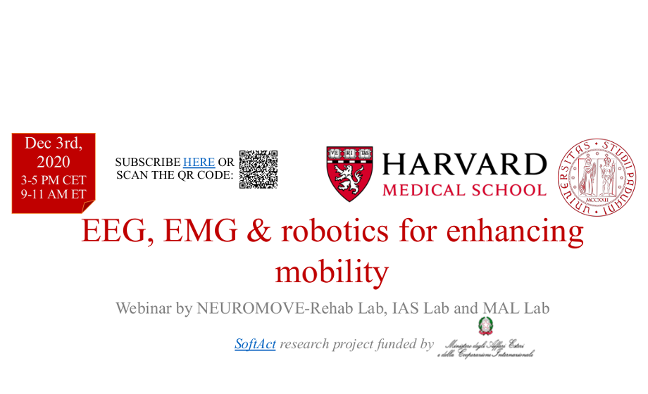
EEG, EMG & robotics for enhancing mobility – Dec. 3rd
When: December 3rd, 2020 – 3:00 – 5:00 PM CET ( 09:00 – 11:00 AM ET )
Where: Zoom Webinar
Webinar by NEUROMOVE-Rehab Lab, IAS Lab and MAL Lab
Please fill the form to subscribe and receive the link to connect a few days before the Webinar.
PNC Brain Day – Sept. 25
When: September 25th, 2020
Where: Aula Ippolito Nievo – Cortile Antico, Palazzo del Bo – Padova
The Padua Neuroscience Center is a unique multidisciplinary environment that integrates many different scientific backgrounds with the common effort to understand the brain mechanisms. In this scenario, sharing scientific ideas and technical expertise represents a crucial and fundamental step to establish collaborative networks and to draft powerful scientific plans.
The PNC Brain Day 2020 is the opportunity for the PIs:
- To get to know each other in a series of short seminars and round tables;
- To contribute to the identification of cross-platform research lines characterizing the PNC scientific plan;
- To render PNC a unique and competitive research unit.
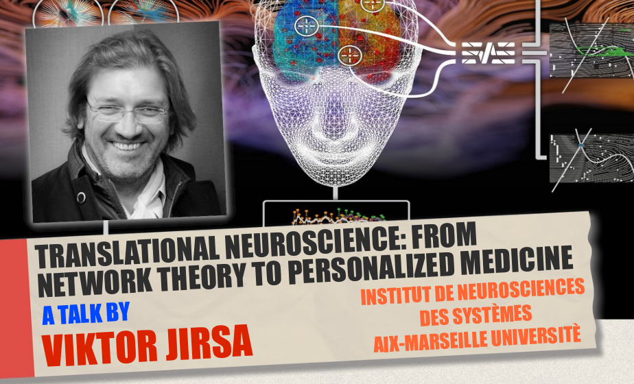
Translational Neuroscience: From Network Theory To Personalized Medicine
by prof. Viktor Jirsa, Institut de Neurosciences, Aix-Marseille Universitè
When: June 18th, 2020 – 3:00 pm
Where: Zoom meeting
Abstract: Over the past decade we have demonstrated that the fusion of subject-specific structural information of the human brain with mathematical dynamic models allows building biologically realistic brain network models, which have a predictive value, beyond the explanatory power of each approach independently. Here we illustrate the workflow along the example of epilepsy: we reconstruct personalized connectivity matrices of human epileptic patients using Diffusion Tensor weighted Imaging (DTI).
Biophysical investigation of the molecular pathogenesis of CMT1X neuropathy
by prof. Mario Bortolozzi, Physics and Astronomy Dept., Padova
When: June 4th, 2020 – 3:00 pm
Where: Zoom meeting
Abstract: Mutations of connexin 32 (Cx32) protein cause the X-linked form of Charcot–Marie–Tooth disease (CMT1X), a demyelinating peripheral neuropathy for which there is no cure. A growing body of evidence indicates that ATP release through Cx32 hemichannels in Schwann cells could be critical for nerve myelination, but it is unknown if CMT1X mutations alter the physiological mechanism that controls Cx32 hemichannel opening and ATP release.
Our study uncovered a link between CMT1X and Cx32 hemichannel dysfunction, suggesting a candidate peptide for treating the disease caused by the R220X mutation of Cx32. The investigation was carried out by a combination of in vitro fluorescence optical microscopy combined with patch clamp and in silico numerical simulations.
Visuomotor control and visual place learning in flies
by prof. Aram Megighian, Dep. of Biomedical Science, Padova
When: May 28th, 2020 – 3:00 pm
Where: Zoom meeting
Abstract: Navigation plays a key role in organisms adaptive behavior. An adequate response to environmental stimuli, is fundamental for supporting food search, social interactions and mating, all of them step physiological mechanisms from the evolutionary point of view.
The lecture will talk about visuomotor responses and place learning studies in flies made in our and other laboratories combining sophisticated quantitative behavioral techniques, fly genetic tools and optogenetics.
A Rational Framework for Studying Neurorehabilitation Interventions
by prof. Nick Ward, Institute of Neurology, UCL Queen Square
When: May 21th, 2020 – 2:30 pm
Where: Zoom meeting
Abstract: Stroke is the most common cause of neurological disability in the world. In the UK alone, there are more people living with the consequences of stroke than with dementia (1.2M vs 0.85M) with an estimated annual cost of £26B. Stroke is still considered a single incident disease with most resources targeted to the first few hours, days or weeks after onset.
Multitasking reveals impaired spatial awareness after stroke
by prof. Mario Bonato, Dep. of General Psychology, Padova
When: May 14th, 2020 – 3:00 pm
Where: Zoom meeting
Abstract: In everyday life contexts sometimes we manage to attend multiple sources of information without particular effort. Sometimes, however, performing two or more tasks together becomes very difficult, like for instance if we have to drive a car in a foggy day while paying attention to a debate on the radio. In these conditions our attention is loaded and we perform what is called “multitasking”.
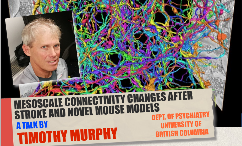
Mesoscale connectivity changes after stroke and novel mouse models
by prof. Timothy Murphy, Dept. of Psychiatry, University of British Columbia
When: May 7th, 2020 – 3:00 pm
Where: Zoom meeting
Abstract: New approaches to real-time assessment and closed-loop feedback based on behavioral features or brain activity will be discussed in the lecture that are designed to optimize stroke recovery interventions in mice for insight into better approaches for human recovery.
The Primary Motor Cortex Seen by a Neurosurgeon: an Anatomical and Functional Appraisal
by Consultant Neurosurgeon PhD. Francesco Vergani, King’s College Hospital, London
When: February 20th, 2020 – 3:00 pm
Where: VIMM Seminar Hall
Abstract: Knowledge of the anatomical and functional relationship between brain tumours and surrounding cortical and subcortical structures is essential in neuro-oncology when planning overall treatment and surgical approach. This is particularly true for tumours in close relationship
to the primary motor cortex and the corticospinal tract (CST), where surgery carries the risk of inducing a permanent motor deficit.
The present review focuses on different aspects of the motor network.
Bridging Neuroscience and Neuroimaging Research to Clinical Practice in Anorexia Nervosa
by prof. Angela Favaro, Dept. of Neuroscience, University of Padova
When: February 6th, 2020 – 3:00 pm
Where: VIMM Seminar Hall
Abstract: Research in the field of neuroimaging, connectomics and neuropsychology is growing in the field of eating disorders.
In this presentation, I will review the recent advances of neuroscience research conducted by our group of research with a particular attention to those aspects that have direct or indirect clinical implications.
Functional Alignment of fMRI Exploiting Prior Information
by prof. Livio Finos, Dept. of Developmental Psychology and Socialisation, University of Padova
When: January 30th, 2020 – 3:00 pm
Where: VIMM Seminar Hall
Abstract: Multi-subject functional Magnetic Resonance Image (fMRI) studies are critical to test the validity of findings across subjects. However, the anatomical and functional structure varies across subjects, hence the image alignment is a fundamental step. One anatomical alignment is the Talairach Atlas, thus, it doesn’t account for functional topography. For that, Haxby et al. (2011) developed a functional approach called Hyperalignment, using sequential Procrustes orthogonal transformations. The inter-subject classification of functional response is improved. However, any constraint isn’t imposed to the transformation, losing results interpretability.
In this presentation, functional connectivity-related phenotypes associated with the risk for the disorders, their modulation by genetic variation and treatment will be discussed.
Imaging and Stimulating Adaptive Brain Plasticity
by prof. Heidi Johansen-Berg, Wellcome Centre for Integrative Neuroimaging, University of Oxford
When: January 23rd, 2020 – 3:00 pm
Where: VIMM Seminar Hall
Abstract: Animal studies show that the adult brain shows remarkable plasticity in response to learning or recovery from injury. Non-invasive brain imaging techniques can be used to detect systems-level structural and functional plasticity in the human brain.
This talk will focus on how brain imaging has allowed us to monitor healthy brains learning new motor skills, to assess how brains recover after damage, such as stroke, and how they adapt to change, such as limb amputation.
Dynamics of Brain networks in Psychosis: Implications for Diagnosis and Treatment
by prof. Fabio Sambataro, Dept. of Neuroscience, University of Padova
When: January 16th, 2020 – 3:00 pm
Where: VIMM Seminar Hall
Abstract: Psychoses are the most severe and devastating psychiatric disorders that cut through the classical nosological categories and include schizophrenia spectrum disorders as well as affective disorders. Genetic and environmental factors have been associated with their etiology, but their pathophysiology is still unknown. Neuroimaging studies have investigated structural and functional changes associated with the risk for these disorders along with their treatment response.
In this presentation, functional connectivity-related phenotypes associated with the risk for the disorders, their modulation by genetic variation and treatment will be discussed.
A Note on the Use of Topic Modeling and Deep Learning to Advance Information Retrieval
by prof. Massimo Melucci, Dept. of Information Engineering, University of Padova
When: January 9th, 2020 – 3:00 pm
Where: VIMM Seminar Hall
Abstract: Information Retrieval (IR) is the complex of theories, models, and technologies aiming to retrieve relevant information to user’s information needs. IR has recently made significant advances in understanding the content of multimedia documents and user queries.
In this talk, I’ll illustrate some noticeable advances, in particular, how topic modeling helps understand content and how the use of deep learning helps overcome some obstacles.
Multilayer network maps of functional human brain
Structure-function relationship in large-scale systems: Brain (and spine) MRI
A talk by Claudia A. M. Gandini Wheeler-Kingshott, University College London.
When: June 28th, 2018
The importance of central-peripheral communication: a neurovisceral integration perspective
May 17th, 2018: “The importance of central-peripheral communication: a neurovisceral integration perspective”
A talk by Prof. Julian F. Thayer, Ohio State University, OH, USA.
Brain networks and neuropsychological disorders in stroke
May 31st, 2018: “Brain networks and neuropsychological disorders in stroke”
A talk by Dr. Antonello Baldassarre: IRCCS Istituto Neurologico Mediterraneo – Neuromed, Italy.
Advances in Placebo Effects
May 7th, 2018: “Advances in Placebo Effects”
A talk by Prof. Irving Kirsch, Harvard Medical School, Beth Israel Deaconess Medical Center, MA, USA.
Detection costs and Identification benefits for cued objects: From peripheral cueing to semantic congruency
May 24th, 2018: “Detection costs and Identification benefits for cued objects: From peripheral cueing to semantic congruency”
A talk by Prof. Juan Lupiañez Castillo, Universidad de Granada, Spain
A computational model of shared fine-scale structure in high-dimensional cortical information spaces
April 12nd, 2018: “A computational model of shared fine-scale structure in high-dimensional cortical information spaces”
A talk by Prof. James Haxby, Dartmouth College, NH, USA.
Detection costs and Identification benefits for cued objects: From peripheral cueing to semantic congruency
May 24th, 2018: “Detection costs and Identification benefits for cued objects: From peripheral cueing to semantic congruency.”
A talk by Juan Lupiañez Castillo, Universidad de Granada.
Optical dissection of the neural circuits underlying sensory perception in the mouse cortex
March 29th, 2018: “Optical dissection of the neural circuits underlying sensory perception in the mouse cortex”
A talk by Prof. Tommaso Fellin, Istituto Italiano di Tecnologia (GE).
Talks on Imaging and Behavior
February 27th, 2018:
“Thalamic spindles in control of sleep and memory consolidation.”
A talk by Prof. Hee-Sup Shin Center for Cognition and Sociality, Institute for Basic Science, Daejon, Korea.
Exploring epilepsy with structural (MR) and functional (e.g. PET) imaging – and what hybrid PET-MR might tell us in addition
February 15th, 2018: “Exploring epilepsy with structural (MR) and functional (e.g. PET) imaging – and what hybrid PET-MR might tell us in addition”
A talk by Prof. Alexander Hammers, King’s College London- St Thomas’ Hospital.
Measuring and manipulating the brain’s intrinsic dynamics
December 6th, 2017: “Measuring and manipulating the brain’s intrinsic dynamics”
A talk by Dr. Christopher M. Lewis, Laboratory of Neural Circuit Dynamics in the Brain Research Institute of the University of Zürich.
Cosa possiamo imparare da neuroimmagini e elettrofisiologia sul disturbo dell’attenzione e iperattività (ADHD)?
November 23rd, 2017: “Cosa possiamo imparare da neuroimmagini e elettrofisiologia sul disturbo dell’attenzione e iperattività (ADHD)?”
A talk by Mario Liotti, docente presso il Dipartimento di Psicologia dello sviluppo e della socializzazione dell’Università degli Studi di Padova.
From vision to navigation: a journey across mouse cortex
A talk by Matteo Carandini, GlaxoSmithKline/Fight for sight Professor of Visual Neuroscience, University College of London.
When: October 26th, 2017
Individual differences in the neural substrates that support factors underlying executive functions
A talk by Marie T. Banich, Professor of Psychology at the University of Colorado Boulder.
When: October 3rd, 2017
Introduction to Resting State fMRI Processing: theory and practices
by Nicholas Metcalf, B.S. Bionformaticist and Data Analyst at Washington University in St. Louis
The course will be held on the following dates:
September 18, 19, 20, 25 and 26th, 2017.
The following concepts and steps in the analysis will be discussed:
- Introduction to fMRI data formats, volume & surface visualization, and anatomy registration
- fMRI registration, distortion correction, and resampling in volume and to surfaces
- fMRI noise removal, temporal filtering and masking
- Overview of the quality control techniques
- How to perform simple statistical analysis
